National Institutes of Health/National Institute Of Allergy and Infectious Diseases (NIH/NIAID)
1R01-AI141465
United States
Citation
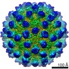
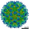
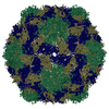
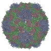
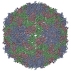
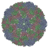
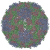
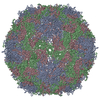
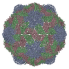
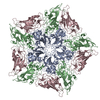
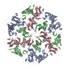
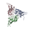
Journal: J Virol / Year: 2021 Title: Multiple Signals in the Gut Contract the Mouse Norovirus Capsid To Block Antibody Binding While Enhancing Receptor Affinity. Authors: Alexis N Williams / Michael B Sherman / Hong Q Smith / Stefan Taube / B Montgomery Pettitt / Christiane E Wobus / Thomas J Smith / Abstract: Human norovirus is the leading cause of gastroenteritis worldwide, with no approved vaccine or antiviral treatment to mitigate infection. These plus-strand RNA viruses have T = 3 icosahedral ...Human norovirus is the leading cause of gastroenteritis worldwide, with no approved vaccine or antiviral treatment to mitigate infection. These plus-strand RNA viruses have T = 3 icosahedral protein capsids with 90 pronounced protruding (P) domain dimers, to which antibodies and cellular receptors bind. We previously demonstrated that bile binding to the capsid of mouse norovirus (MNV) causes several major conformational changes; the entire P domain rotates by ∼90° and contracts onto the shell, the P domain dimers rotate about each other, and the structural equilibrium of the epitopes at the top of the P domain shifts toward the closed conformation, which favors receptor binding while blocking antibody binding. Here, we demonstrate that MNV undergoes reversible conformational changes at pH 5.0 that are nearly identical to those observed when bile binds. Notably, at low pH or when metals bind, a cluster of acidic resides in the G'-H' loop interact and distort the G'-H' loop, and this may drive C'-D' loop movement toward the closed conformation. Enzyme-linked immunosorbent assays with infectious virus particles at low pH or in the presence of metals demonstrated that all tested antibodies do not bind to this contracted form, akin to what was observed with the MNV-bile complex. Therefore, low pH, cationic metals, and bile salts are physiological triggers in the gut for P domain contraction and structural rearrangement, which synergistically prime the virus for receptor binding while blocking antibody binding. The protruding domains on the calicivirus capsids are recognized by cell receptors and antibodies. We demonstrated that MNV P domains are highly mobile, and bile causes contraction onto the shell surface while allosterically blocking antibody binding. We present the near-atomic cryo-electron microscopy structures of infectious MNV at pH 5.0 and pH 7.5. Surprisingly, low pH is sufficient to cause the same conformational changes as when bile binds. A cluster of acidic residues on the G'-H' loop were most likely involved in the pH effects. These residues also bound divalent cations and had the same conformation as observed here at pH 5. Binding assays demonstrated that low pH and metals block antibody binding, and thus the G'-H' loop might be driving the conformational changes. Therefore, low pH, cationic metals, and bile salts in the gut synergistically prime the virus for receptor binding while blocking antibody binding.
In the structure databanks used in Yorodumi, some data are registered as the other names, "COVID-19 virus" and "2019-nCoV". Here are the details of the virus and the list of structure data.
Jan 31, 2019. EMDB accession codes are about to change! (news from PDBe EMDB page)
EMDB accession codes are about to change! (news from PDBe EMDB page)
The allocation of 4 digits for EMDB accession codes will soon come to an end. Whilst these codes will remain in use, new EMDB accession codes will include an additional digit and will expand incrementally as the available range of codes is exhausted. The current 4-digit format prefixed with “EMD-” (i.e. EMD-XXXX) will advance to a 5-digit format (i.e. EMD-XXXXX), and so on. It is currently estimated that the 4-digit codes will be depleted around Spring 2019, at which point the 5-digit format will come into force.
The EM Navigator/Yorodumi systems omit the EMD- prefix.
Related info.:Q: What is EMD? / ID/Accession-code notation in Yorodumi/EM Navigator
Yorodumi is a browser for structure data from EMDB, PDB, SASBDB, etc.
This page is also the successor to EM Navigator detail page, and also detail information page/front-end page for Omokage search.
The word "yorodu" (or yorozu) is an old Japanese word meaning "ten thousand". "mi" (miru) is to see.
Related info.:EMDB / PDB / SASBDB / Comparison of 3 databanks / Yorodumi Search / Aug 31, 2016. New EM Navigator & Yorodumi / Yorodumi Papers / Jmol/JSmol / Function and homology information / Changes in new EM Navigator and Yorodumi
 Movie
Movie Controller


 Open data
 Basic information
 Components
 Keywords
 Function and homology information
 Murine norovirus 1
 Authors
 United States, 1items
  Citation
 Structure visualization
 Downloads & links
 Other downloads
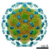


 PDBj
PDBj
 Assembly
 Sample preparation
 Electron microscopy imaging
 FIELD EMISSION GUN / Accelerating voltage: 300 kV / Illumination mode: FLOOD BEAM
 Processing