 Movie
Movie Controller


+ Open data

- Basic information
| Entry | Database: PDB / ID: 7mry | ||||||
|---|---|---|---|---|---|---|---|
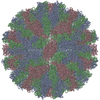
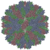
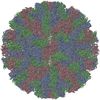
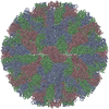
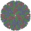
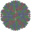
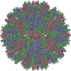
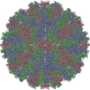
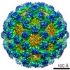
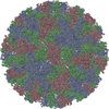
| Title | Norovirus T=3 GII.4 HOV VLP | ||||||
 Components Components | VP1 | ||||||
 Keywords Keywords | VIRUS / T=3 Capsid GII.4 HOV Norovirus | ||||||
| Function / homology | Calicivirus coat protein C-terminal / Calicivirus coat protein C-terminal / Calicivirus coat protein / Calicivirus coat protein / virion component / Picornavirus/Calicivirus coat protein / Viral coat protein subunit / host cell cytoplasm / VP1 Function and homology information Function and homology information | ||||||
| Biological species |  Norovirus GII.4 Norovirus GII.4 | ||||||
| Method | ELECTRON MICROSCOPY / single particle reconstruction / cryo EM / Resolution: 3.8 Å | ||||||
 Authors Authors | Salmen, W. / Hu, L. / Prasad, B. | ||||||
| Funding support |  United States, 1items United States, 1items
| ||||||
 Citation Citation | Journal: Nat Commun / Year: 2022 Title: Atomic structure of the predominant GII.4 human norovirus capsid reveals novel stability and plasticity. Authors: Liya Hu / Wilhelm Salmen / Rong Chen / Yi Zhou / Frederick Neill / James E Crowe / Robert L Atmar / Mary K Estes / B V Venkataram Prasad / Abstract: Human noroviruses (HuNoVs) cause sporadic and epidemic viral gastroenteritis worldwide. The GII.4 variants are responsible for most HuNoV infections, and GII.4 virus-like particles (VLPs) are being ...Human noroviruses (HuNoVs) cause sporadic and epidemic viral gastroenteritis worldwide. The GII.4 variants are responsible for most HuNoV infections, and GII.4 virus-like particles (VLPs) are being used in vaccine development. The atomic structure of the GII.4 capsid in the native T = 3 state has not been determined. Here we present the GII.4 VLP structure with T = 3 symmetry determined using X-ray crystallography and cryo-EM at 3.0 Å and 3.8 Å resolution, respectively, which reveals unanticipated novel features. A novel aspect in the crystal structure determined without imposing icosahedral symmetry is the remarkable adaptability of the capsid protein VP1 driven by the flexible hinge between the shell and the protruding domains. In both crystal and cryo-EM structures, VP1 adopts a stable conformation with the protruding domain resting on the shell domain, in contrast to the 'rising' conformation observed in recent cryo-EM structures of other GII.4 VLPs. Our studies further revealed that the resting state of VP1 dimer is stabilized by a divalent ion, and chelation using EDTA increases capsid diameter, exposing new hydrophobic and antigenic sites and suggesting a transition to the rising conformation. These novel insights into GII.4 capsid structure, stability, and antigen presentation may be useful for ongoing vaccine development. | ||||||
| History |
|
- Structure visualization
| Movie |
Movie viewer |
|---|---|
| Structure viewer | Molecule: MolmilJmol/JSmol |
- Downloads & links
-Download
| PDBx/mmCIF format | 7mry.cif.gz | 266.4 KB | Display | PDBx/mmCIF format |
|---|---|---|---|---|
| PDB format | pdb7mry.ent.gz | 208.1 KB | Display | PDB format |
| PDBx/mmJSON format | 7mry.json.gz | Tree view | PDBx/mmJSON format | |
| Others |  Other downloads Other downloads |
-Validation report
| Arichive directory | https://data.pdbj.org/pub/pdb/validation_reports/mr/7mryftp://data.pdbj.org/pub/pdb/validation_reports/mr/7mry | HTTPS FTP |
|---|
-Related structure data
| Related structure data |  23960MC M: map data used to model this data C: citing same article ( |
|---|---|
| Similar structure data |

-Links
 PDBj
PDBj
- Assembly
| Deposited unit | 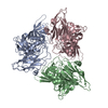
|
|---|---|
| 1 | x 60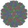
|
| 2 |
|
| 3 | x 5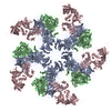
|
| 4 | x 6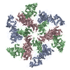
|
| 5 | 
|
| Symmetry | Point symmetry: (Schoenflies symbol: I (icosahedral)) |
-Components
| #1: Protein | Mass: 58760.895 Da / Num. of mol.: 3 Source method: isolated from a genetically manipulated source Source: (gene. exp.) Norovirus GII.4 / Production host:  Trichoplusia ni (cabbage looper) / References: UniProt: A9YYE4 Trichoplusia ni (cabbage looper) / References: UniProt: A9YYE4#2: Water | ChemComp-HOH / |  Mass: 18.015 Da / Num. of mol.: 5 / Source method: isolated from a natural source / Formula: H2O Mass: 18.015 Da / Num. of mol.: 5 / Source method: isolated from a natural source / Formula: H2O |
|---|
-Experimental details
-Experiment
| Experiment | Method: ELECTRON MICROSCOPY |
|---|---|
| EM experiment | Aggregation state: PARTICLE / 3D reconstruction method: single particle reconstruction |
- Sample preparation
| Component | Name: Norovirus GII.4 / Type: VIRUS / Entity ID: #1 / Source: RECOMBINANT |
|---|---|
| Source (natural) | Organism: Norovirus GII.4 |
| Source (recombinant) | Organism: Trichoplusia ni (cabbage looper) |
| Details of virus | Empty: YES / Enveloped: NO / Isolate: STRAIN / Type: VIRUS-LIKE PARTICLE |
| Buffer solution | pH: 6 |
| Specimen | Conc.: 0.96 mg/ml / Embedding applied: NO / Shadowing applied: NO / Staining applied: NO / Vitrification applied: YES |
| Vitrification | Cryogen name: ETHANE |
- Electron microscopy imaging
| Microscopy | Model: JEOL 3200FSC |
|---|---|
| Electron gun | Electron source:  FIELD EMISSION GUN / Accelerating voltage: 300 kV / Illumination mode: SPOT SCAN FIELD EMISSION GUN / Accelerating voltage: 300 kV / Illumination mode: SPOT SCAN |
| Electron lens | Mode: BRIGHT FIELD |
| Image recording | Average exposure time: 10 sec. / Electron dose: 55.96 e/Å2 / Film or detector model: GATAN K2 SUMMIT (4k x 4k) |
- Processing
| Software |
| ||||||||||||||||||||||||
|---|---|---|---|---|---|---|---|---|---|---|---|---|---|---|---|---|---|---|---|---|---|---|---|---|---|
| EM software |
| ||||||||||||||||||||||||
| CTF correction | Type: PHASE FLIPPING AND AMPLITUDE CORRECTION | ||||||||||||||||||||||||
| 3D reconstruction | Resolution: 3.8 Å / Resolution method: FSC 0.143 CUT-OFF / Num. of particles: 51913 Details: Using Cryosparc GSFSC, resolution calculated: No Mask = 4.2 A, Spherical = 4 A, Loose = 3.9 A, Tight = 3.8 A, corrected = 3.8 A Symmetry type: POINT | ||||||||||||||||||||||||
| Refinement | Cross valid method: NONE Stereochemistry target values: GeoStd + Monomer Library + CDL v1.2 | ||||||||||||||||||||||||
| Displacement parameters | Biso mean: 76.67 Å2 | ||||||||||||||||||||||||
| Refine LS restraints |
|
