 Movie
Movie Controller

[English] 日本語
 Yorodumi
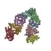
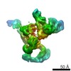
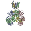
- PDB-7m7i: 6-Deoxyerythronolide B synthase (DEBS) module 1 in complex with a... -
+ Open data

- Basic information
| Entry | Database: PDB / ID: 7m7i | |||||||||||||||||||||||||||
|---|---|---|---|---|---|---|---|---|---|---|---|---|---|---|---|---|---|---|---|---|---|---|---|---|---|---|---|---|
| Title | 6-Deoxyerythronolide B synthase (DEBS) module 1 in complex with antibody fragment 1B2 (TE-free) | |||||||||||||||||||||||||||
 Components Components |
| |||||||||||||||||||||||||||
 Keywords Keywords | BIOSYNTHETIC PROTEIN/IMMUNE SYSTEM / polyketide synthase / antibody fragment / BIOSYNTHETIC PROTEIN-IMMUNE SYSTEM complex | |||||||||||||||||||||||||||
| Function / homology |  Function and homology information Function and homology information6-deoxyerythronolide-B synthase / fatty acid synthase activity / phosphopantetheine binding / 3-oxoacyl-[acyl-carrier-protein] synthase activity / antibiotic biosynthetic process / fatty acid biosynthetic process Similarity search - Function | |||||||||||||||||||||||||||
| Biological species |  Saccharopolyspora erythraea (bacteria) Saccharopolyspora erythraea (bacteria) Homo sapiens (human) Homo sapiens (human) | |||||||||||||||||||||||||||
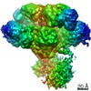
| Method | ELECTRON MICROSCOPY / single particle reconstruction / cryo EM / Resolution: 3.4 Å | |||||||||||||||||||||||||||
 Authors Authors | Cogan, D.P. / Zhang, K. / Chiu, W. / Khosla, C. | |||||||||||||||||||||||||||
| Funding support |  United States, 1items United States, 1items
| |||||||||||||||||||||||||||
 Citation Citation | Journal: Science / Year: 2021 Title: Mapping the catalytic conformations of an assembly-line polyketide synthase module. Authors: Dillon P Cogan / Kaiming Zhang / Xiuyuan Li / Shanshan Li / Grigore D Pintilie / Soung-Hun Roh / Charles S Craik / Wah Chiu / Chaitan Khosla /   Abstract: Assembly-line polyketide synthases, such as the 6-deoxyerythronolide B synthase (DEBS), are large enzyme factories prized for their ability to produce specific and complex polyketide products. By ...Assembly-line polyketide synthases, such as the 6-deoxyerythronolide B synthase (DEBS), are large enzyme factories prized for their ability to produce specific and complex polyketide products. By channeling protein-tethered substrates across multiple active sites in a defined linear sequence, these enzymes facilitate programmed small-molecule syntheses that could theoretically be harnessed to access countless polyketide product structures. Using cryogenic electron microscopy to study DEBS module 1, we present a structural model describing this substrate-channeling phenomenon. Our 3.2- to 4.3-angstrom-resolution structures of the intact module reveal key domain-domain interfaces and highlight an unexpected module asymmetry. We also present the structure of a product-bound module that shines light on a recently described “turnstile” mechanism for transient gating of active sites along the assembly line. | |||||||||||||||||||||||||||
| History |
|
- Structure visualization
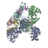
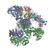
| Movie |
Movie viewer |
|---|---|
| Structure viewer | Molecule: MolmilJmol/JSmol |
- Downloads & links
-Download
| PDBx/mmCIF format | 7m7i.cif.gz | 547.3 KB | Display | PDBx/mmCIF format |
|---|---|---|---|---|
| PDB format | pdb7m7i.ent.gz | 436.7 KB | Display | PDB format |
| PDBx/mmJSON format | 7m7i.json.gz | Tree view | PDBx/mmJSON format | |
| Others |  Other downloads Other downloads |
-Validation report
| Summary document | 7m7i_validation.pdf.gz | 986.1 KB | Display | wwPDB validaton report |
|---|---|---|---|---|
| Full document | 7m7i_full_validation.pdf.gz | 1 MB | Display | |
| Data in XML | 7m7i_validation.xml.gz | 88.4 KB | Display | |
| Data in CIF | 7m7i_validation.cif.gz | 135.7 KB | Display | |
| Arichive directory | https://data.pdbj.org/pub/pdb/validation_reports/m7/7m7iftp://data.pdbj.org/pub/pdb/validation_reports/m7/7m7i | HTTPS FTP |
-Related structure data
| Related structure data |  23714MC  7m7eC 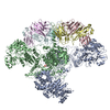 7m7fC  7m7gC  7m7hC  7m7jC M: map data used to model this data C: citing same article ( |
|---|---|
| Similar structure data |


-Links
 PDBj
PDBj


- Assembly
| Deposited unit | 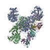
|
|---|---|
| 1 |
|
-Components
| #1: Protein | Mass: 167782.750 Da / Num. of mol.: 2 / Fragment: UNP residues 557-2010,3463-3545 Source method: isolated from a genetically manipulated source Source: (gene. exp.) Saccharopolyspora erythraea (bacteria) / Gene: eryAI / Production host: #2: Antibody | Mass: 26447.611 Da / Num. of mol.: 2 Source method: isolated from a genetically manipulated source Source: (gene. exp.) Saccharopolyspora erythraea (bacteria) / Production host: #3: Antibody | Mass: 25715.832 Da / Num. of mol.: 2 Source method: isolated from a genetically manipulated source Source: (gene. exp.) Homo sapiens (human) / Production host: #4: Chemical | ChemComp-PN7 / |   Mass: 358.348 Da / Num. of mol.: 1 Mass: 358.348 Da / Num. of mol.: 1Source method: isolated from a genetically manipulated source Formula: C11H23N2O7PS Has ligand of interest | Y | Has protein modification | Y | |
|---|
-Experimental details
-Experiment
| Experiment | Method: ELECTRON MICROSCOPY |
|---|---|
| EM experiment | Aggregation state: PARTICLE / 3D reconstruction method: single particle reconstruction |
- Sample preparation
| Component | Name: Complex between DEBS (3)M1(2) and antibody fragment 1B2 Type: COMPLEX / Entity ID: #1-#3 / Source: MULTIPLE SOURCES | ||||||||||||||||||||
|---|---|---|---|---|---|---|---|---|---|---|---|---|---|---|---|---|---|---|---|---|---|
| Molecular weight | Value: 0.44 MDa / Experimental value: NO | ||||||||||||||||||||
| Source (natural) | Organism: Saccharopolyspora erythraea (bacteria) | ||||||||||||||||||||
| Source (recombinant) | Organism: | ||||||||||||||||||||
| Buffer solution | pH: 7.2 | ||||||||||||||||||||
| Buffer component |
| ||||||||||||||||||||
| Specimen | Conc.: 10 mg/ml / Embedding applied: NO / Shadowing applied: NO / Staining applied: NO / Vitrification applied: YES | ||||||||||||||||||||
| Vitrification | Instrument: FEI VITROBOT MARK IV / Cryogen name: ETHANE / Humidity: 100 % / Chamber temperature: 277.15 K |
- Electron microscopy imaging
| Experimental equipment |  Model: Titan Krios / Image courtesy: FEI Company |
|---|---|
| Microscopy | Model: FEI TITAN KRIOS |
| Electron gun | Electron source:  FIELD EMISSION GUN / Accelerating voltage: 300 kV / Illumination mode: OTHER FIELD EMISSION GUN / Accelerating voltage: 300 kV / Illumination mode: OTHER |
| Electron lens | Mode: BRIGHT FIELD |
| Specimen holder | Cryogen: NITROGEN / Specimen holder model: FEI TITAN KRIOS AUTOGRID HOLDER |
| Image recording | Average exposure time: 8.5 sec. / Electron dose: 50 e/Å2 / Film or detector model: FEI FALCON IV (4k x 4k) / Num. of real images: 8104 |
- Processing
| Software | Name: PHENIX / Version: 1.19_4092: / Classification: refinement | ||||||||||||||||||||||||
|---|---|---|---|---|---|---|---|---|---|---|---|---|---|---|---|---|---|---|---|---|---|---|---|---|---|
| EM software |
| ||||||||||||||||||||||||
| CTF correction | Type: PHASE FLIPPING AND AMPLITUDE CORRECTION | ||||||||||||||||||||||||
| Particle selection | Num. of particles selected: 466815 | ||||||||||||||||||||||||
| Symmetry | Point symmetry: C1 (asymmetric) | ||||||||||||||||||||||||
| 3D reconstruction | Resolution: 3.4 Å / Resolution method: FSC 0.143 CUT-OFF / Num. of particles: 69566 / Symmetry type: POINT | ||||||||||||||||||||||||
| Refine LS restraints |
|
