 Movie
Movie Controller

[English] 日本語
 Yorodumi
- PDB-7lh2: Structure of human prestin in the presence of sodium salicylate a... -
+ Open data

- Basic information
| Entry | Database: PDB / ID: 7lh2 | |||||||||||||||||||||||||||
|---|---|---|---|---|---|---|---|---|---|---|---|---|---|---|---|---|---|---|---|---|---|---|---|---|---|---|---|---|
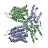
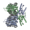
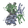
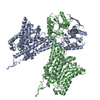
| Title | Structure of human prestin in the presence of sodium salicylate and sodium sulfate | |||||||||||||||||||||||||||
 Components Components | Prestin | |||||||||||||||||||||||||||
 Keywords Keywords | MEMBRANE PROTEIN / transporter family / lipid interaction / high resolution | |||||||||||||||||||||||||||
| Function / homology |  Function and homology information Function and homology informationlateral wall of outer hair cell / oxalate transport / fructose transmembrane transport / response to salicylic acid / oxalate transmembrane transporter activity / sulfate transmembrane transporter activity / secondary active sulfate transmembrane transporter activity / sulfate transmembrane transport / response to thyroid hormone / response to potassium ion ...lateral wall of outer hair cell / oxalate transport / fructose transmembrane transport / response to salicylic acid / oxalate transmembrane transporter activity / sulfate transmembrane transporter activity / secondary active sulfate transmembrane transporter activity / sulfate transmembrane transport / response to thyroid hormone / response to potassium ion / negative regulation of monoatomic ion transmembrane transport / chloride:bicarbonate antiporter activity / response to salt / response to auditory stimulus / bicarbonate transport / Sensory processing of sound by outer hair cells of the cochlea / positive regulation of cell motility / chloride transport / spectrin binding / cochlea development / lateral plasma membrane / positive regulation of cell size / response to ischemia / chloride transmembrane transport / regulation of membrane potential / sensory perception of sound / regulation of cell shape / basolateral plasma membrane / response to xenobiotic stimulus / protein homodimerization activity / plasma membrane Similarity search - Function | |||||||||||||||||||||||||||
| Biological species |  Homo sapiens (human) Homo sapiens (human) | |||||||||||||||||||||||||||
| Method | ELECTRON MICROSCOPY / single particle reconstruction / cryo EM / Resolution: 3.43 Å | |||||||||||||||||||||||||||
 Authors Authors | Ge, J. / Gouaux, E. | |||||||||||||||||||||||||||
| Funding support |  United States, 1items United States, 1items
| |||||||||||||||||||||||||||
 Citation Citation | Journal: Cell / Year: 2021 Title: Molecular mechanism of prestin electromotive signal amplification. Authors: Jingpeng Ge / Johannes Elferich / Sepehr Dehghani-Ghahnaviyeh / Zhiyu Zhao / Marc Meadows / Henrique von Gersdorff / Emad Tajkhorshid / Eric Gouaux / Abstract: Hearing involves two fundamental processes: mechano-electrical transduction and signal amplification. Despite decades of studies, the molecular bases for both remain elusive. Here, we show how ...Hearing involves two fundamental processes: mechano-electrical transduction and signal amplification. Despite decades of studies, the molecular bases for both remain elusive. Here, we show how prestin, the electromotive molecule of outer hair cells (OHCs) that senses both voltage and membrane tension, mediates signal amplification by coupling conformational changes to alterations in membrane surface area. Cryoelectron microscopy (cryo-EM) structures of human prestin bound with chloride or salicylate at a common "anion site" adopt contracted or expanded states, respectively. Prestin is ensconced within a perimeter of well-ordered lipids, through which it induces dramatic deformation in the membrane and couples protein conformational changes to the bulk membrane. Together with computational studies, we illustrate how the anion site is allosterically coupled to changes in the transmembrane domain cross-sectional area and the surrounding membrane. These studies provide insight into OHC electromotility by providing a structure-based mechanism of the membrane motor prestin. | |||||||||||||||||||||||||||
| History |
|
- Structure visualization
| Movie |
Movie viewer |
|---|---|
| Structure viewer | Molecule: MolmilJmol/JSmol |
- Downloads & links
-Download
| PDBx/mmCIF format | 7lh2.cif.gz | 240 KB | Display | PDBx/mmCIF format |
|---|---|---|---|---|
| PDB format | pdb7lh2.ent.gz | 192 KB | Display | PDB format |
| PDBx/mmJSON format | 7lh2.json.gz | Tree view | PDBx/mmJSON format | |
| Others |  Other downloads Other downloads |
-Validation report
| Arichive directory | https://data.pdbj.org/pub/pdb/validation_reports/lh/7lh2ftp://data.pdbj.org/pub/pdb/validation_reports/lh/7lh2 | HTTPS FTP |
|---|
-Related structure data
| Related structure data |  23334MC  7lguC  7lgwC  7lh3C M: map data used to model this data C: citing same article ( |
|---|---|
| Similar structure data |


-Links
 PDBj
PDBj


- Assembly
| Deposited unit | 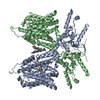
|
|---|---|
| 1 |
|
-Components
| #1: Protein | Mass: 82064.211 Da / Num. of mol.: 2 Source method: isolated from a genetically manipulated source Source: (gene. exp.) Homo sapiens (human) / Gene: SLC26A5, PRES / Production host: Homo sapiens (human) / References: UniProt: P58743#2: Chemical |   Mass: 138.121 Da / Num. of mol.: 2 / Source method: obtained synthetically / Formula: C7H6O3 / Feature type: SUBJECT OF INVESTIGATION Mass: 138.121 Da / Num. of mol.: 2 / Source method: obtained synthetically / Formula: C7H6O3 / Feature type: SUBJECT OF INVESTIGATION#3: Chemical |   Mass: 386.654 Da / Num. of mol.: 2 / Source method: obtained synthetically / Formula: C27H46O / Feature type: SUBJECT OF INVESTIGATION Mass: 386.654 Da / Num. of mol.: 2 / Source method: obtained synthetically / Formula: C27H46O / Feature type: SUBJECT OF INVESTIGATIONHas ligand of interest | Y | Has protein modification | N | |
|---|
-Experimental details
-Experiment
| Experiment | Method: ELECTRON MICROSCOPY |
|---|---|
| EM experiment | Aggregation state: PARTICLE / 3D reconstruction method: single particle reconstruction |
- Sample preparation
| Component | Name: Protein structure bound with substrates and lipids / Type: COMPLEX / Entity ID: #1 / Source: RECOMBINANT |
|---|---|
| Source (natural) | Organism: Homo sapiens (human) |
| Source (recombinant) | Organism: Homo sapiens (human) |
| Buffer solution | pH: 8 |
| Specimen | Embedding applied: NO / Shadowing applied: NO / Staining applied: NO / Vitrification applied: YES |
| Vitrification | Instrument: FEI VITROBOT MARK IV / Cryogen name: ETHANE / Humidity: 95 % / Chamber temperature: 281 K |
- Electron microscopy imaging
| Experimental equipment |  Model: Titan Krios / Image courtesy: FEI Company |
|---|---|
| Microscopy | Model: FEI TITAN KRIOS |
| Electron gun | Electron source:  FIELD EMISSION GUN / Accelerating voltage: 300 kV / Illumination mode: FLOOD BEAM FIELD EMISSION GUN / Accelerating voltage: 300 kV / Illumination mode: FLOOD BEAM |
| Electron lens | Mode: BRIGHT FIELD |
| Image recording | Electron dose: 50 e/Å2 / Film or detector model: GATAN K3 (6k x 4k) |
- Processing
| Software | Name: PHENIX / Version: 1.18.2_3874: / Classification: refinement | ||||||||||||||||||||||||
|---|---|---|---|---|---|---|---|---|---|---|---|---|---|---|---|---|---|---|---|---|---|---|---|---|---|
| EM software | Name: PHENIX / Category: model refinement | ||||||||||||||||||||||||
| CTF correction | Type: PHASE FLIPPING AND AMPLITUDE CORRECTION | ||||||||||||||||||||||||
| 3D reconstruction | Resolution: 3.43 Å / Resolution method: FSC 0.143 CUT-OFF / Num. of particles: 57000 / Symmetry type: POINT | ||||||||||||||||||||||||
| Refine LS restraints |
|
