 ムービー
ムービー コントローラー


+ データを開く

- 基本情報
基本情報
| 登録情報 | データベース: PDB / ID: 7ktt | ||||||
|---|---|---|---|---|---|---|---|
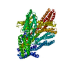
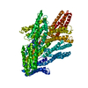
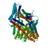
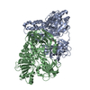
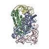
| タイトル | Cryogenic electron microscopy model of full-length human metavinculin | ||||||
 要素 要素 | metavinculin | ||||||
 キーワード キーワード | CELL ADHESION / actin / adaptor protein / cadherin / cancer / catenin / cell junction / cell migration / cell signaling / focal adhesions / heart failure / inositol phospholipid / integrin / plasma membrane / skeletal muscle / smooth muscle | ||||||
| 機能・相同性 |  機能・相同性情報 機能・相同性情報regulation of protein localization to adherens junction / podosome ring / outer dense plaque of desmosome / inner dense plaque of desmosome / terminal web / cell-substrate junction / epithelial cell-cell adhesion / zonula adherens / dystroglycan binding / alpha-catenin binding ...regulation of protein localization to adherens junction / podosome ring / outer dense plaque of desmosome / inner dense plaque of desmosome / terminal web / cell-substrate junction / epithelial cell-cell adhesion / zonula adherens / dystroglycan binding / alpha-catenin binding / fascia adherens / cell-cell contact zone / adherens junction assembly / apical junction assembly / costamere / regulation of establishment of endothelial barrier / axon extension / protein localization to cell surface / lamellipodium assembly / regulation of focal adhesion assembly / maintenance of blood-brain barrier / brush border / Smooth Muscle Contraction / negative regulation of cell migration / cell-matrix adhesion / cell projection / morphogenesis of an epithelium / adherens junction / Signaling by high-kinase activity BRAF mutants / MAP2K and MAPK activation / sarcolemma / beta-catenin binding / platelet aggregation / specific granule lumen / Signaling by RAF1 mutants / Signaling by moderate kinase activity BRAF mutants / Paradoxical activation of RAF signaling by kinase inactive BRAF / Signaling downstream of RAS mutants / Signaling by BRAF and RAF1 fusions / cell-cell junction / extracellular vesicle / Signaling by ALK fusions and activated point mutants / Platelet degranulation / actin binding / secretory granule lumen / ficolin-1-rich granule lumen / molecular adaptor activity / cytoskeleton / cell adhesion / cadherin binding / membrane raft / focal adhesion / ubiquitin protein ligase binding / Neutrophil degranulation / structural molecule activity / protein-containing complex / extracellular exosome / extracellular region / plasma membrane / cytoplasm / cytosol 類似検索 - 分子機能 | ||||||
| 生物種 |  Homo sapiens (ヒト) Homo sapiens (ヒト) | ||||||
| 手法 | 電子顕微鏡法 / 単粒子再構成法 / クライオ電子顕微鏡法 / 解像度: 4.17 Å | ||||||
 データ登録者 データ登録者 | Izard, T. / Rangarajan, E.S. | ||||||
 引用 引用 | ジャーナル: Int J Mol Sci / 年: 2021 タイトル: The Cryogenic Electron Microscopy Structure of the Cell Adhesion Regulator Metavinculin Reveals an Isoform-Specific Kinked Helix in Its Cytoskeleton Binding Domain. 著者: Erumbi S Rangarajan / Tina Izard /  要旨: Vinculin and its heart-specific splice variant metavinculin are key regulators of cell adhesion processes. These membrane-bound cytoskeletal proteins regulate the cell shape by binding to several ...Vinculin and its heart-specific splice variant metavinculin are key regulators of cell adhesion processes. These membrane-bound cytoskeletal proteins regulate the cell shape by binding to several other proteins at cell-cell and cell-matrix junctions. Vinculin and metavinculin link integrin adhesion molecules to the filamentous actin network. Loss of both proteins prevents cell adhesion and cell spreading and reduces the formation of stress fibers, focal adhesions, or lamellipodia extensions. The binding of talin at cell-matrix junctions or of α-catenin at cell-cell junctions activates vinculin and metavinculin by releasing their autoinhibitory head-tail interaction. Once activated, vinculin and metavinculin bind F-actin via their five-helix bundle tail domains. Unlike vinculin, metavinculin has a 68-amino-acid insertion before the second α-helix of this five-helix F-actin-binding domain. Here, we present the full-length cryogenic electron microscopy structure of metavinculin that captures the dynamics of its individual domains and unveiled a hallmark structural feature, namely a kinked isoform-specific α-helix in its F-actin-binding domain. Our identified conformational landscape of metavinculin suggests a structural priming mechanism that is consistent with the cell adhesion functions of metavinculin in response to mechanical and cellular cues. Our findings expand our understanding of metavinculin function in the heart with implications for the etiologies of cardiomyopathies. | ||||||
| 履歴 |
|
- 構造の表示
構造の表示
| ムービー |
ムービービューア |
|---|---|
| 構造ビューア | 分子: MolmilJmol/JSmol |
- ダウンロードとリンク
-ダウンロード
| PDBx/mmCIF形式 | 7ktt.cif.gz | 179.9 KB | 表示 | PDBx/mmCIF形式 |
|---|---|---|---|---|
| PDB形式 | pdb7ktt.ent.gz | 139.7 KB | 表示 | PDB形式 |
| PDBx/mmJSON形式 | 7ktt.json.gz | ツリー表示 | PDBx/mmJSON形式 | |
| その他 |  その他のダウンロード その他のダウンロード |
-検証レポート
| 文書・要旨 | 7ktt_validation.pdf.gz | 809 KB | 表示 | wwPDB検証レポート |
|---|---|---|---|---|
| 文書・詳細版 | 7ktt_full_validation.pdf.gz | 824.5 KB | 表示 | |
| XML形式データ | 7ktt_validation.xml.gz | 32.3 KB | 表示 | |
| CIF形式データ | 7ktt_validation.cif.gz | 47.7 KB | 表示 | |
| アーカイブディレクトリ | https://data.pdbj.org/pub/pdb/validation_reports/kt/7kttftp://data.pdbj.org/pub/pdb/validation_reports/kt/7ktt | HTTPS FTP |
-関連構造データ
| 関連構造データ |  23029MC  7ktuC  7ktvC  7ktwC M: このデータのモデリングに利用したマップデータ C: 同じ文献を引用 ( |
|---|---|
| 類似構造データ | |
| 電子顕微鏡画像生データ | EMPIAR-11029 (タイトル: Cryogenic electron microscopy structure of full length human meta vinculin Data size: 744.8 Data #1: Raw movies of human metavinculin [micrographs - multiframe]) |


-リンク
 PDBj
PDBj


- 集合体
集合体
| 登録構造単位 | 
|
|---|---|
| 1 |
|
-要素
| #1: タンパク質 | 分子量: 125061.500 Da / 分子数: 1 / 由来タイプ: 組換発現 / 由来: (組換発現) Homo sapiens (ヒト) / 遺伝子: VCL / 発現宿主:  |
|---|
-実験情報
-実験
| 実験 | 手法: 電子顕微鏡法 |
|---|---|
| EM実験 | 試料の集合状態: PARTICLE / 3次元再構成法: 単粒子再構成法 |
- 試料調製
試料調製
| 構成要素 | 名称: metavinculin / タイプ: COMPLEX / 詳細: Full-length / Entity ID: all / 由来: RECOMBINANT |
|---|---|
| 由来(天然) | 生物種: Homo sapiens (ヒト) |
| 由来(組換発現) | 生物種: |
| 緩衝液 | pH: 8 / 詳細: 20 mM Tris, pH 8.0, 150 mM NaCl, 0.2 mM TCEP |
| 試料 | 濃度: 0.5 mg/ml / 包埋: NO / シャドウイング: NO / 染色: NO / 凍結: YES |
| 試料支持 | グリッドの材料: COPPER / グリッドのサイズ: 300 divisions/in. / グリッドのタイプ: Quantifoil R1.2/1.3 |
| 急速凍結 | 装置: LEICA EM GP / 凍結剤: ETHANE / 湿度: 95 % / 凍結前の試料温度: 277 K |
- 電子顕微鏡撮影
| 実験機器 |  モデル: Titan Krios / 画像提供: FEI Company |
|---|---|
| 顕微鏡 | モデル: FEI TITAN KRIOS |
| 電子銃 | 電子線源:  FIELD EMISSION GUN / 加速電圧: 300 kV / 照射モード: SPOT SCAN FIELD EMISSION GUN / 加速電圧: 300 kV / 照射モード: SPOT SCAN |
| 電子レンズ | モード: BRIGHT FIELD / 倍率(公称値): 105000 X / 最大 デフォーカス(公称値): 1400 nm / 最小 デフォーカス(公称値): 1400 nm / Cs: 2.7 mm / アライメント法: ZEMLIN TABLEAU |
| 試料ホルダ | 凍結剤: NITROGEN 試料ホルダーモデル: FEI TITAN KRIOS AUTOGRID HOLDER |
| 撮影 | 平均露光時間: 1.6 sec. / 電子線照射量: 67.92 e/Å2 / フィルム・検出器のモデル: GATAN K3 (6k x 4k) / 撮影したグリッド数: 1 / 実像数: 2998 |
| 電子光学装置 | エネルギーフィルタースリット幅: 20 eV |
- 解析
解析
| ソフトウェア | 名称: PHENIX / バージョン: 1.13_2998: / 分類: 精密化 | ||||||||||||||||||||||||||||
|---|---|---|---|---|---|---|---|---|---|---|---|---|---|---|---|---|---|---|---|---|---|---|---|---|---|---|---|---|---|
| EMソフトウェア |
| ||||||||||||||||||||||||||||
| CTF補正 | タイプ: PHASE FLIPPING AND AMPLITUDE CORRECTION | ||||||||||||||||||||||||||||
| 対称性 | 点対称性: C1 (非対称) | ||||||||||||||||||||||||||||
| 3次元再構成 | 解像度: 4.17 Å / 解像度の算出法: FSC 0.143 CUT-OFF / 粒子像の数: 1303504 / アルゴリズム: FOURIER SPACE / 対称性のタイプ: POINT | ||||||||||||||||||||||||||||
| 原子モデル構築 | プロトコル: RIGID BODY FIT | ||||||||||||||||||||||||||||
| 拘束条件 |
|
