 Movie
Movie Controller

[English] 日本語
 Yorodumi
- PDB-7ktw: Cryogenic electron microscopy model of full-length human metavinc... -
+ Open data

- Basic information
| Entry | Database: PDB / ID: 7ktw | |||||||||||||||||||||
|---|---|---|---|---|---|---|---|---|---|---|---|---|---|---|---|---|---|---|---|---|---|---|
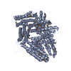
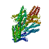
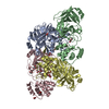
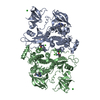
| Title | Cryogenic electron microscopy model of full-length human metavinculin H1'-parallel conformation 2 | |||||||||||||||||||||
 Components Components | metavinculin | |||||||||||||||||||||
 Keywords Keywords | CELL ADHESION / actin / adaptor protein / cadherin / cancer / catenin / cell junction / cell migration / cell signaling / focal adhesions / heart failure / inositol phospholipid / integrin / plasma membrane / skeletal muscle / smooth muscle | |||||||||||||||||||||
| Function / homology |  Function and homology information Function and homology informationregulation of protein localization to adherens junction / outer dense plaque of desmosome / inner dense plaque of desmosome / podosome ring / terminal web / cell-substrate junction / epithelial cell-cell adhesion / zonula adherens / fascia adherens / dystroglycan binding ...regulation of protein localization to adherens junction / outer dense plaque of desmosome / inner dense plaque of desmosome / podosome ring / terminal web / cell-substrate junction / epithelial cell-cell adhesion / zonula adherens / fascia adherens / dystroglycan binding / alpha-catenin binding / cell-cell contact zone / apical junction assembly / costamere / regulation of establishment of endothelial barrier / Regulation of CDH1 Function / protein localization to cell surface / adherens junction assembly / lamellipodium assembly / regulation of focal adhesion assembly / maintenance of blood-brain barrier / brush border / Smooth Muscle Contraction / cell-matrix adhesion / negative regulation of cell migration / Turbulent (oscillatory, disturbed) flow shear stress activates signaling by PIEZO1 and integrins in endothelial cells / morphogenesis of an epithelium / adherens junction / Signaling by high-kinase activity BRAF mutants / MAP2K and MAPK activation / sarcolemma / platelet aggregation / beta-catenin binding / specific granule lumen / Signaling by RAF1 mutants / cell-cell junction / Signaling by moderate kinase activity BRAF mutants / Paradoxical activation of RAF signaling by kinase inactive BRAF / Signaling downstream of RAS mutants / Signaling by BRAF and RAF1 fusions / Signaling by ALK fusions and activated point mutants / Platelet degranulation / extracellular vesicle / actin binding / secretory granule lumen / High laminar flow shear stress activates signaling by PIEZO1 and PECAM1:CDH5:KDR in endothelial cells / molecular adaptor activity / ficolin-1-rich granule lumen / cytoskeleton / cell adhesion / cadherin binding / membrane raft / focal adhesion / ubiquitin protein ligase binding / Neutrophil degranulation / perinuclear region of cytoplasm / structural molecule activity / protein-containing complex / extracellular exosome / extracellular region / plasma membrane / cytosol / cytoplasm Similarity search - Function | |||||||||||||||||||||
| Biological species |  Homo sapiens (human) Homo sapiens (human) | |||||||||||||||||||||
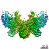
| Method | ELECTRON MICROSCOPY / single particle reconstruction / cryo EM / Resolution: 4.27 Å | |||||||||||||||||||||
 Authors Authors | Izard, T. / Rangarajan, E.S. | |||||||||||||||||||||
| Funding support |  United States, 1items United States, 1items
| |||||||||||||||||||||
 Citation Citation | Journal: Int J Mol Sci / Year: 2021 Title: The Cryogenic Electron Microscopy Structure of the Cell Adhesion Regulator Metavinculin Reveals an Isoform-Specific Kinked Helix in Its Cytoskeleton Binding Domain. Authors: Erumbi S Rangarajan / Tina Izard / Abstract: Vinculin and its heart-specific splice variant metavinculin are key regulators of cell adhesion processes. These membrane-bound cytoskeletal proteins regulate the cell shape by binding to several ...Vinculin and its heart-specific splice variant metavinculin are key regulators of cell adhesion processes. These membrane-bound cytoskeletal proteins regulate the cell shape by binding to several other proteins at cell-cell and cell-matrix junctions. Vinculin and metavinculin link integrin adhesion molecules to the filamentous actin network. Loss of both proteins prevents cell adhesion and cell spreading and reduces the formation of stress fibers, focal adhesions, or lamellipodia extensions. The binding of talin at cell-matrix junctions or of α-catenin at cell-cell junctions activates vinculin and metavinculin by releasing their autoinhibitory head-tail interaction. Once activated, vinculin and metavinculin bind F-actin via their five-helix bundle tail domains. Unlike vinculin, metavinculin has a 68-amino-acid insertion before the second α-helix of this five-helix F-actin-binding domain. Here, we present the full-length cryogenic electron microscopy structure of metavinculin that captures the dynamics of its individual domains and unveiled a hallmark structural feature, namely a kinked isoform-specific α-helix in its F-actin-binding domain. Our identified conformational landscape of metavinculin suggests a structural priming mechanism that is consistent with the cell adhesion functions of metavinculin in response to mechanical and cellular cues. Our findings expand our understanding of metavinculin function in the heart with implications for the etiologies of cardiomyopathies. | |||||||||||||||||||||
| History |
|
- Structure visualization
| Movie |
Movie viewer |
|---|---|
| Structure viewer | Molecule: MolmilJmol/JSmol |
- Downloads & links
-Download
| PDBx/mmCIF format | 7ktw.cif.gz | 180.6 KB | Display | PDBx/mmCIF format |
|---|---|---|---|---|
| PDB format | pdb7ktw.ent.gz | 139.8 KB | Display | PDB format |
| PDBx/mmJSON format | 7ktw.json.gz | Tree view | PDBx/mmJSON format | |
| Others |  Other downloads Other downloads |
-Validation report
| Arichive directory | https://data.pdbj.org/pub/pdb/validation_reports/kt/7ktwftp://data.pdbj.org/pub/pdb/validation_reports/kt/7ktw | HTTPS FTP |
|---|
-Related structure data
| Related structure data |  23032MC  7kttC  7ktuC  7ktvC M: map data used to model this data C: citing same article ( |
|---|---|
| Similar structure data | |
| EM raw data | EMPIAR-11029 (Title: Cryogenic electron microscopy structure of full length human meta vinculin Data size: 744.8 Data #1: Raw movies of human metavinculin [micrographs - multiframe]) |


-Links
 PDBj
PDBj


- Assembly
| Deposited unit | 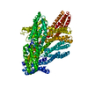
|
|---|---|
| 1 |
|
-Components
| #1: Protein | Mass: 125061.500 Da / Num. of mol.: 1 Source method: isolated from a genetically manipulated source Source: (gene. exp.) Homo sapiens (human) / Gene: VCL / Production host:  |
|---|---|
| Has protein modification | N |
-Experimental details
-Experiment
| Experiment | Method: ELECTRON MICROSCOPY |
|---|---|
| EM experiment | Aggregation state: PARTICLE / 3D reconstruction method: single particle reconstruction |
- Sample preparation
| Component | Name: metavinculin / Type: COMPLEX / Entity ID: all / Source: RECOMBINANT |
|---|---|
| Source (natural) | Organism: Homo sapiens (human) |
| Source (recombinant) | Organism: |
| Buffer solution | pH: 8 / Details: 20 mM Tris, pH 8.0, 150 mM NaCl, 0.2 mM TCEP |
| Specimen | Conc.: 0.5 mg/ml / Embedding applied: NO / Shadowing applied: NO / Staining applied: NO / Vitrification applied: YES |
| Specimen support | Grid material: COPPER / Grid mesh size: 300 divisions/in. / Grid type: Quantifoil R1.2/1.3 |
| Vitrification | Instrument: LEICA EM GP / Cryogen name: ETHANE / Humidity: 95 % / Chamber temperature: 277 K |
- Electron microscopy imaging
| Experimental equipment |  Model: Titan Krios / Image courtesy: FEI Company |
|---|---|
| Microscopy | Model: FEI TITAN KRIOS |
| Electron gun | Electron source:  FIELD EMISSION GUN / Accelerating voltage: 300 kV / Illumination mode: SPOT SCAN FIELD EMISSION GUN / Accelerating voltage: 300 kV / Illumination mode: SPOT SCAN |
| Electron lens | Mode: BRIGHT FIELD / Nominal magnification: 105000 X / Nominal defocus max: 1400 nm / Nominal defocus min: 1400 nm / Cs: 2.7 mm / Alignment procedure: ZEMLIN TABLEAU |
| Specimen holder | Cryogen: NITROGEN / Specimen holder model: FEI TITAN KRIOS AUTOGRID HOLDER |
| Image recording | Average exposure time: 1.6 sec. / Electron dose: 67.92 e/Å2 / Film or detector model: GATAN K3 (6k x 4k) / Num. of grids imaged: 1 / Num. of real images: 2998 |
| EM imaging optics | Energyfilter slit width: 20 eV |
- Processing
| Software | Name: PHENIX / Version: 1.13_2998: / Classification: refinement | ||||||||||||||||||||||||||||||||
|---|---|---|---|---|---|---|---|---|---|---|---|---|---|---|---|---|---|---|---|---|---|---|---|---|---|---|---|---|---|---|---|---|---|
| EM software |
| ||||||||||||||||||||||||||||||||
| CTF correction | Type: PHASE FLIPPING AND AMPLITUDE CORRECTION | ||||||||||||||||||||||||||||||||
| Symmetry | Point symmetry: C1 (asymmetric) | ||||||||||||||||||||||||||||||||
| 3D reconstruction | Resolution: 4.27 Å / Resolution method: FSC 0.143 CUT-OFF / Num. of particles: 366802 / Symmetry type: POINT | ||||||||||||||||||||||||||||||||
| Atomic model building | Protocol: RIGID BODY FIT | ||||||||||||||||||||||||||||||||
| Refine LS restraints |
|
