 Movie
Movie Controller

[English] 日本語
 Yorodumi
- PDB-7kf5: Cryo-electron microscopy structure of the heavy metal efflux pump... -
+ Open data

- Basic information
| Entry | Database: PDB / ID: 7kf5 | ||||||
|---|---|---|---|---|---|---|---|
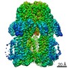
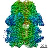
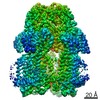
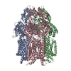
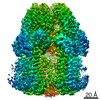
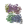
| Title | Cryo-electron microscopy structure of the heavy metal efflux pump CusA in the symmetric closed state | ||||||
 Components Components | Cation efflux system protein CusA | ||||||
 Keywords Keywords | MEMBRANE PROTEIN / TRANSPORT PROTEIN / efflux / pump / heavy metal. copper / silver / closed / open / transport | ||||||
| Function / homology |  Function and homology information Function and homology informationsilver ion transport / silver ion transmembrane transporter activity / plasma membrane copper ion transport / copper ion transmembrane transport / response to silver ion / silver ion transmembrane transport / copper ion transmembrane transporter activity / copper ion export / detoxification of copper ion / response to copper ion ...silver ion transport / silver ion transmembrane transporter activity / plasma membrane copper ion transport / copper ion transmembrane transport / response to silver ion / silver ion transmembrane transport / copper ion transmembrane transporter activity / copper ion export / detoxification of copper ion / response to copper ion / xenobiotic transmembrane transporter activity / intracellular copper ion homeostasis / response to toxic substance / copper ion binding / membrane / plasma membrane Similarity search - Function | ||||||
| Biological species |  | ||||||
| Method | ELECTRON MICROSCOPY / single particle reconstruction / cryo EM / Resolution: 3.2 Å | ||||||
 Authors Authors | Moseng, M.A. | ||||||
| Funding support |  United States, 1items United States, 1items
| ||||||
 Citation Citation | Journal: mBio / Year: 2021 Title: Cryo-EM Structures of CusA Reveal a Mechanism of Metal-Ion Export. Authors: Mitchell A Moseng / Meinan Lyu / Tanadet Pipatpolkai / Przemyslaw Glaza / Corey C Emerson / Phoebe L Stewart / Phillip J Stansfeld / Edward W Yu /  Abstract: Gram-negative bacteria utilize the resistance-nodulation-cell division (RND) superfamily of efflux pumps to expel a variety of toxic compounds from the cell. The CusA membrane protein, which ...Gram-negative bacteria utilize the resistance-nodulation-cell division (RND) superfamily of efflux pumps to expel a variety of toxic compounds from the cell. The CusA membrane protein, which recognizes and extrudes biocidal Cu(I) and Ag(I) ions, belongs to the heavy-metal efflux (HME) subfamily of RND efflux pumps. We here report four structures of the trimeric CusA heavy-metal efflux pump in the presence of Cu(I) using single-particle cryo-electron microscopy (cryo-EM). We discover that different CusA protomers within the trimer are able to bind Cu(I) ions simultaneously. Our structural data combined with molecular dynamics (MD) simulations allow us to propose a mechanism for ion transport where each CusA protomer functions independently within the trimer. The bacterial RND superfamily of efflux pumps mediate resistance to a variety of biocides, including Cu(I) and Ag(I) ions. Here we report four cryo-EM structures of the trimeric CusA pump in the presence of Cu(I). Combined with MD simulations, our data indicate that each CusA protomer within the trimer recognizes and extrudes Cu(I) independently. | ||||||
| History |
|
- Structure visualization
| Movie |
Movie viewer |
|---|---|
| Structure viewer | Molecule: MolmilJmol/JSmol |
- Downloads & links
-Download
| PDBx/mmCIF format | 7kf5.cif.gz | 989.6 KB | Display | PDBx/mmCIF format |
|---|---|---|---|---|
| PDB format | pdb7kf5.ent.gz | 817.6 KB | Display | PDB format |
| PDBx/mmJSON format | 7kf5.json.gz | Tree view | PDBx/mmJSON format | |
| Others |  Other downloads Other downloads |
-Validation report
| Arichive directory | https://data.pdbj.org/pub/pdb/validation_reports/kf/7kf5ftp://data.pdbj.org/pub/pdb/validation_reports/kf/7kf5 | HTTPS FTP |
|---|
-Related structure data
| Related structure data | 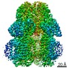 22843MC 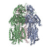 7kf6C 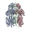 7kf7C 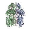 7kf8C M: map data used to model this data C: citing same article ( |
|---|---|
| Similar structure data |
-Links
 PDBj
PDBj
- Assembly
| Deposited unit | 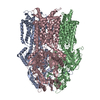
|
|---|---|
| 1 |
|
-Components
| #1: Protein | Mass: 115833.945 Da / Num. of mol.: 3 Source method: isolated from a genetically manipulated source Source: (gene. exp.) |
|---|
-Experimental details
-Experiment
| Experiment | Method: ELECTRON MICROSCOPY |
|---|---|
| EM experiment | Aggregation state: PARTICLE / 3D reconstruction method: single particle reconstruction |
- Sample preparation
| Component | Name: CusA in symmetric closed extrusion form / Type: COMPLEX / Entity ID: all / Source: RECOMBINANT |
|---|---|
| Molecular weight | Experimental value: YES |
| Source (natural) | Organism: |
| Source (recombinant) | Organism: |
| Buffer solution | pH: 7 |
| Specimen | Conc.: 1 mg/ml / Embedding applied: NO / Shadowing applied: NO / Staining applied: NO / Vitrification applied: YES |
| Specimen support | Grid material: COPPER / Grid mesh size: 300 divisions/in. / Grid type: Quantifoil R1.2/1.3 |
| Vitrification | Instrument: FEI VITROBOT MARK I / Cryogen name: ETHANE / Humidity: 100 % / Chamber temperature: 277.5 K |
- Electron microscopy imaging
| Experimental equipment |  Model: Titan Krios / Image courtesy: FEI Company |
|---|---|
| Microscopy | Model: FEI TITAN KRIOS |
| Electron gun | Electron source:  FIELD EMISSION GUN / Accelerating voltage: 300 kV / Illumination mode: OTHER FIELD EMISSION GUN / Accelerating voltage: 300 kV / Illumination mode: OTHER |
| Electron lens | Mode: BRIGHT FIELD |
| Image recording | Electron dose: 40 e/Å2 / Film or detector model: GATAN K2 SUMMIT (4k x 4k) |
- Processing
| CTF correction | Type: NONE |
|---|---|
| 3D reconstruction | Resolution: 3.2 Å / Resolution method: FSC 0.143 CUT-OFF / Num. of particles: 13304 / Symmetry type: POINT |
