 Movie
Movie Controller

[English] 日本語
 Yorodumi
- PDB-7jti: Interphotoreceptor retinoid-binding protein (IRBP) in complex wit... -
+ Open data

- Basic information
| Entry | Database: PDB / ID: 7jti | |||||||||||||||
|---|---|---|---|---|---|---|---|---|---|---|---|---|---|---|---|---|
| Title | Interphotoreceptor retinoid-binding protein (IRBP) in complex with a monoclonal antibody (F3F5 mAb5) | |||||||||||||||
 Components Components |
| |||||||||||||||
 Keywords Keywords | TRANSPORT PROTEIN/IMMUNE SYSTEM / IRBP / retinoid / Interphotoreceptor retinoid-binding protein / Antibody / TRANSPORT PROTEIN-IMMUNE SYSTEM complex | |||||||||||||||
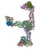
| Function / homology |  Function and homology information Function and homology informationcone matrix sheath / interphotoreceptor matrix / all-trans-retinol binding / oleic acid binding / retinal binding / retinol binding / serine-type peptidase activity / proteolysis Similarity search - Function | |||||||||||||||
| Biological species |  | |||||||||||||||
| Method | ELECTRON MICROSCOPY / single particle reconstruction / cryo EM / Resolution: 7.4 Å | |||||||||||||||
 Authors Authors | Sears, A.E. / Albiez, S. / Gulati, S. / Wang, B. / Kiser, P. / Kovacik, L. / Engel, A. / Stahlberg, H. / Palczewski, K. | |||||||||||||||
| Funding support |  United States, United States,  Switzerland, 4items Switzerland, 4items
| |||||||||||||||
 Citation Citation | Journal: FASEB J / Year: 2020 Title: Single particle cryo-EM of the complex between interphotoreceptor retinoid-binding protein and a monoclonal antibody. Authors: Avery E Sears / Stefan Albiez / Sahil Gulati / Benlian Wang / Philip Kiser / Lubomir Kovacik / Andreas Engel / Henning Stahlberg / Krzysztof Palczewski / Abstract: Interphotoreceptor retinoid-binding protein (IRBP) is a highly expressed protein secreted by rod and cone photoreceptors that has major roles in photoreceptor homeostasis as well as retinoid and ...Interphotoreceptor retinoid-binding protein (IRBP) is a highly expressed protein secreted by rod and cone photoreceptors that has major roles in photoreceptor homeostasis as well as retinoid and polyunsaturated fatty acid transport between the neural retina and retinal pigment epithelium. Despite two crystal structures reported on fragments of IRBP and decades of research, the overall structure of IRBP and function within the visual cycle remain unsolved. Here, we studied the structure of native bovine IRBP in complex with a monoclonal antibody (mAb5) by cryo-electron microscopy, revealing the tertiary and quaternary structure at sufficient resolution to clearly identify the complex components. Complementary mass spectrometry experiments revealed the structure and locations of N-linked carbohydrate post-translational modifications. This work provides insight into the structure of IRBP, displaying an elongated, flexible three-dimensional architecture not seen among other retinoid-binding proteins. This work is the first step in elucidation of the function of this enigmatic protein. | |||||||||||||||
| History |
|
- Structure visualization
| Movie |
Movie viewer |
|---|---|
| Structure viewer | Molecule: MolmilJmol/JSmol |
- Downloads & links
-Download
| PDBx/mmCIF format | 7jti.cif.gz | 299.9 KB | Display | PDBx/mmCIF format |
|---|---|---|---|---|
| PDB format | pdb7jti.ent.gz | 236.1 KB | Display | PDB format |
| PDBx/mmJSON format | 7jti.json.gz | Tree view | PDBx/mmJSON format | |
| Others |  Other downloads Other downloads |
-Validation report
| Arichive directory | https://data.pdbj.org/pub/pdb/validation_reports/jt/7jtiftp://data.pdbj.org/pub/pdb/validation_reports/jt/7jti | HTTPS FTP |
|---|
-Related structure data
| Related structure data | 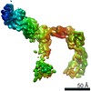 22474MC M: map data used to model this data C: citing same article ( |
|---|---|
| Similar structure data | |
| EM raw data | EMPIAR-10388 (Title: Single particle cryo-EM of the complex between interphotoreceptor retinoid-binding protein and a monoclonal antibody Data size: 729.9 Data #1: Unaligned multi-frame micrographs of IRBP and antibody [micrographs - multiframe]) |

-Links
 PDBj
PDBj

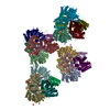
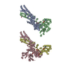
- Assembly
| Deposited unit | 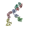
|
|---|---|
| 1 |
|
-Components
-Retinol-binding protein ... , 4 types, 4 molecules ABCD
| #3: Protein | Mass: 33267.301 Da / Num. of mol.: 1 / Fragment: Module 1 (UNP residues 329-631) / Source method: isolated from a natural source / Source: (natural) |
|---|---|
| #4: Protein | Mass: 33386.637 Da / Num. of mol.: 1 / Fragment: Module 2 / Source method: isolated from a natural source / Source: (natural) |
| #5: Protein | Mass: 32399.768 Da / Num. of mol.: 1 / Fragment: Module 3 (UNP residues 633-932) / Source method: isolated from a natural source / Source: (natural) |
| #6: Protein | Mass: 32192.508 Da / Num. of mol.: 1 / Fragment: Module 4 (UNP residues 936-1231) / Source method: isolated from a natural source / Source: (natural) |
-Antibody , 2 types, 2 molecules LH
| #1: Antibody | Mass: 22596.021 Da / Num. of mol.: 1 Source method: isolated from a genetically manipulated source Source: (gene. exp.) |
|---|---|
| #2: Antibody | Mass: 23777.783 Da / Num. of mol.: 1 Source method: isolated from a genetically manipulated source Source: (gene. exp.) |
-Details
| Has protein modification | Y |
|---|
-Experimental details
-Experiment
| Experiment | Method: ELECTRON MICROSCOPY |
|---|---|
| EM experiment | Aggregation state: PARTICLE / 3D reconstruction method: single particle reconstruction |
- Sample preparation
| Component |
| |||||||||||||||||||||||||
|---|---|---|---|---|---|---|---|---|---|---|---|---|---|---|---|---|---|---|---|---|---|---|---|---|---|---|
| Molecular weight | Value: 0.193 MDa / Experimental value: YES | |||||||||||||||||||||||||
| Source (natural) |
| |||||||||||||||||||||||||
| Source (recombinant) | Organism: | |||||||||||||||||||||||||
| Buffer solution | pH: 8 | |||||||||||||||||||||||||
| Buffer component |
| |||||||||||||||||||||||||
| Specimen | Conc.: 0.2 mg/ml / Embedding applied: NO / Shadowing applied: NO / Staining applied: NO / Vitrification applied: YES Details: The sample had minor aggregation but was overall monodisperse. | |||||||||||||||||||||||||
| Specimen support | Grid material: COPPER / Grid mesh size: 400 divisions/in. / Grid type: Quantifoil R1.2/1.3 | |||||||||||||||||||||||||
| Vitrification | Instrument: FEI VITROBOT MARK IV / Cryogen name: ETHANE / Humidity: 90 % / Chamber temperature: 277 K / Details: Blot for 3 seconds before plunging. |
- Electron microscopy imaging
| Experimental equipment |  Model: Titan Krios / Image courtesy: FEI Company |
|---|---|
| Microscopy | Model: FEI TITAN KRIOS |
| Electron gun | Electron source:  FIELD EMISSION GUN / Accelerating voltage: 300 kV / Illumination mode: FLOOD BEAM FIELD EMISSION GUN / Accelerating voltage: 300 kV / Illumination mode: FLOOD BEAM |
| Electron lens | Mode: BRIGHT FIELD / Nominal magnification: 60000 X / Nominal defocus max: 2200 nm / Nominal defocus min: 1000 nm / Alignment procedure: BASIC |
| Specimen holder | Cryogen: NITROGEN Specimen holder model: GATAN 910 MULTI-SPECIMEN SINGLE TILT CRYO TRANSFER HOLDER |
| Image recording | Average exposure time: 1 sec. / Electron dose: 80 e/Å2 / Detector mode: SUPER-RESOLUTION / Film or detector model: GATAN K2 QUANTUM (4k x 4k) / Num. of grids imaged: 4 / Num. of real images: 2649 Details: Images were collected in movie mode at 35 frames per second. |
| EM imaging optics | Energyfilter name: GIF Quantum LS / Energyfilter slit width: 20 eV |
| Image scans | Width: 1000 / Height: 1000 / Movie frames/image: 35 |
- Processing
| EM software |
| ||||||||||||||||||||||||||||||||
|---|---|---|---|---|---|---|---|---|---|---|---|---|---|---|---|---|---|---|---|---|---|---|---|---|---|---|---|---|---|---|---|---|---|
| Image processing | Details: The selected images were low-pass filtered and normalized. | ||||||||||||||||||||||||||||||||
| CTF correction | Type: PHASE FLIPPING AND AMPLITUDE CORRECTION | ||||||||||||||||||||||||||||||||
| Particle selection | Num. of particles selected: 281804 Details: Ab initio, cisTEM processing using low-pass filtered reconstruction | ||||||||||||||||||||||||||||||||
| 3D reconstruction | Resolution: 7.4 Å / Resolution method: FSC 0.5 CUT-OFF / Num. of particles: 17900 / Symmetry type: POINT | ||||||||||||||||||||||||||||||||
| Atomic model building | B value: 156 / Protocol: RIGID BODY FIT / Space: REAL / Target criteria: Correlation coefficient | ||||||||||||||||||||||||||||||||
| Atomic model building | PDB-ID: 1J7X Pdb chain-ID: A / Accession code: 1J7X / Source name: PDB / Type: experimental model |

