National Health and Medical Research Council (NHMRC, Australia)
APP1109901
オーストラリア
引用
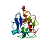
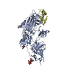
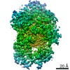
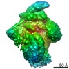
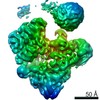
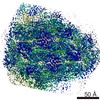
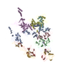
ジャーナル: J Biol Chem / 年: 2021 タイトル: The cryo-EM structure of the endocytic receptor DEC-205. 著者: Benjamin S Gully / Hariprasad Venugopal / Alex J Fulcher / Zhihui Fu / Jessica Li / Felix A Deuss / Carmen Llerena / William R Heath / Mireille H Lahoud / Irina Caminschi / Jamie Rossjohn / Richard Berry / 要旨: DEC-205 (CD205), a member of the macrophage mannose receptor protein family, is the prototypic endocytic receptor of dendritic cells, whose ligands include phosphorothioated cytosine-guanosine ...DEC-205 (CD205), a member of the macrophage mannose receptor protein family, is the prototypic endocytic receptor of dendritic cells, whose ligands include phosphorothioated cytosine-guanosine oligonucleotides, a motif often seen in bacterial or viral DNA. However, despite growing biological and clinical significance, little is known about the structural arrangement of this receptor or any of its family members. Here, we describe the 3.2 Å cryo-EM structure of human DEC-205, thereby illuminating the structure of the mannose receptor protein family. The DEC-205 monomer forms a compact structure comprising two intercalated rings of C-type lectin-like domains, where the N-terminal cysteine-rich and fibronectin domains reside at the central intersection. We establish a pH-dependent oligomerization pathway forming tetrameric DEC-205 using solution-based techniques and ultimately solved the 4.9 Å cryo-EM structure of the DEC-205 tetramer to identify the unfurling of the second lectin ring which enables tetramer formation. Furthermore, we suggest the relevance of this oligomerization pathway within a cellular setting, whereby cytosine-guanosine binding appeared to disrupt this cell-surface oligomer. Accordingly, we provide insight into the structure and oligomeric assembly of the DEC-205 receptor.
 ムービー
ムービー コントローラー


 データを開く
 基本情報
基本情報 要素
要素 キーワード
キーワード 機能・相同性情報
 Homo sapiens (ヒト)
 データ登録者
 オーストラリア, 2件
  引用
引用
 構造の表示
構造の表示 ダウンロードとリンク
 その他のダウンロード

 PDBj
PDBj


 集合体
集合体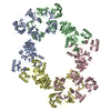
 試料調製
試料調製 電子顕微鏡撮影
 FIELD EMISSION GUN / 加速電圧: 300 kV / 照射モード: FLOOD BEAM
 解析
解析