 Movie
Movie Controller

[English] 日本語
 Yorodumi
- PDB-7jk9: Helical filaments of plant light-dependent protochlorophyllide ox... -
+ Open data

- Basic information
| Entry | Database: PDB / ID: 7jk9 | ||||||||||||
|---|---|---|---|---|---|---|---|---|---|---|---|---|---|
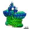
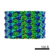
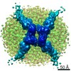
| Title | Helical filaments of plant light-dependent protochlorophyllide oxidoreductase (LPOR) bound to NADPH, Pchlide, and membrane | ||||||||||||
 Components Components | Protochlorophyllide reductase B, chloroplastic | ||||||||||||
 Keywords Keywords | PHOTOSYNTHESIS / reductase / light-activated / ligand-protein complex | ||||||||||||
| Function / homology |  Function and homology information Function and homology informationprotochlorophyllide reductase / protochlorophyllide reductase activity / chloroplast outer membrane / response to ethylene / chlorophyll biosynthetic process / chloroplast thylakoid / chloroplast envelope / chloroplast thylakoid membrane / photosynthesis / chloroplast ...protochlorophyllide reductase / protochlorophyllide reductase activity / chloroplast outer membrane / response to ethylene / chlorophyll biosynthetic process / chloroplast thylakoid / chloroplast envelope / chloroplast thylakoid membrane / photosynthesis / chloroplast / protein domain specific binding / mRNA binding / cytosol Similarity search - Function | ||||||||||||
| Biological species |  | ||||||||||||
| Method | ELECTRON MICROSCOPY / helical reconstruction / cryo EM / Resolution: 3.1 Å | ||||||||||||
 Authors Authors | Nguyen, H.C. / Gabruk, M. / Frost, A. | ||||||||||||
| Funding support |  Poland, Poland,  United States, 3items United States, 3items
| ||||||||||||
 Citation Citation | Journal: Nat Plants / Year: 2021 Title: Photocatalytic LPOR forms helical lattices that shape membranes for chlorophyll synthesis. Authors: Henry C Nguyen / Arthur A Melo / Jerzy Kruk / Adam Frost / Michal Gabruk / Abstract: Chlorophyll biosynthesis, crucial to life on Earth, is tightly regulated because its precursors are phototoxic. In flowering plants, the enzyme light-dependent protochlorophyllide oxidoreductase ...Chlorophyll biosynthesis, crucial to life on Earth, is tightly regulated because its precursors are phototoxic. In flowering plants, the enzyme light-dependent protochlorophyllide oxidoreductase (LPOR) captures photons to catalyse the penultimate reaction: the reduction of a double bond within protochlorophyllide (Pchlide) to generate chlorophyllide (Chlide). In darkness, LPOR oligomerizes to facilitate photon energy transfer and catalysis. However, the complete three-dimensional structure of LPOR, the higher-order architecture of LPOR oligomers and the implications of these self-assembled states for catalysis, including how LPOR positions Pchlide and the co-factor NADPH, remain unknown. Here, we report the atomic structure of LPOR assemblies by electron cryo-microscopy. LPOR polymerizes with its substrates into helical filaments around constricted lipid bilayer tubes. Portions of LPOR and Pchlide insert into the outer membrane leaflet, targeting the product, Chlide, to the membrane for the final reaction site of chlorophyll biosynthesis. In addition to its crucial photocatalytic role, we show that in darkness LPOR filaments directly shape membranes into high-curvature tubules with the spectral properties of the prolamellar body, whose light-triggered disassembly provides lipids for thylakoid assembly. Moreover, our structure of the catalytic site challenges previously proposed reaction mechanisms. Together, our results reveal a new and unexpected synergy between photosynthetic membrane biogenesis and chlorophyll synthesis in plants, orchestrated by LPOR. | ||||||||||||
| History |
|
- Structure visualization
| Movie |
Movie viewer |
|---|---|
| Structure viewer | Molecule: MolmilJmol/JSmol |
- Downloads & links
-Download
| PDBx/mmCIF format | 7jk9.cif.gz | 4 MB | Display | PDBx/mmCIF format |
|---|---|---|---|---|
| PDB format | pdb7jk9.ent.gz | Display | PDB format | |
| PDBx/mmJSON format | 7jk9.json.gz | Tree view | PDBx/mmJSON format | |
| Others |  Other downloads Other downloads |
-Validation report
| Summary document | 7jk9_validation.pdf.gz | 8.8 MB | Display | wwPDB validaton report |
|---|---|---|---|---|
| Full document | 7jk9_full_validation.pdf.gz | 9 MB | Display | |
| Data in XML | 7jk9_validation.xml.gz | 620.1 KB | Display | |
| Data in CIF | 7jk9_validation.cif.gz | 1.3 MB | Display | |
| Arichive directory | https://data.pdbj.org/pub/pdb/validation_reports/jk/7jk9ftp://data.pdbj.org/pub/pdb/validation_reports/jk/7jk9 | HTTPS FTP |
-Related structure data
| Related structure data |  22364MC M: map data used to model this data C: citing same article ( |
|---|---|
| Similar structure data |

-Links
 PDBj
PDBj


- Assembly
| Deposited unit | 
|
|---|---|
| 1 |
|
| Symmetry | Helical symmetry: (Circular symmetry: 2 / Dyad axis: no / N subunits divisor: 1 / Num. of operations: 6 / Rise per n subunits: 43.1 Å / Rotation per n subunits: 50.34 °) |
-Components
| #1: Protein | Mass: 43415.199 Da / Num. of mol.: 40 Source method: isolated from a genetically manipulated source Source: (gene. exp.)  #2: Chemical | ChemComp-NDP /   Mass: 745.421 Da / Num. of mol.: 40 / Source method: obtained synthetically / Formula: C21H30N7O17P3 Mass: 745.421 Da / Num. of mol.: 40 / Source method: obtained synthetically / Formula: C21H30N7O17P3#3: Chemical | ChemComp-PMR /   Mass: 612.957 Da / Num. of mol.: 40 / Source method: obtained synthetically / Formula: C35H32MgN4O5 / Feature type: SUBJECT OF INVESTIGATION Mass: 612.957 Da / Num. of mol.: 40 / Source method: obtained synthetically / Formula: C35H32MgN4O5 / Feature type: SUBJECT OF INVESTIGATION#4: Chemical | ChemComp-LMG /   Mass: 787.158 Da / Num. of mol.: 40 / Source method: obtained synthetically / Formula: C45H86O10 Mass: 787.158 Da / Num. of mol.: 40 / Source method: obtained synthetically / Formula: C45H86O10Has ligand of interest | Y | |
|---|
-Experimental details
-Experiment
| Experiment | Method: ELECTRON MICROSCOPY |
|---|---|
| EM experiment | Aggregation state: FILAMENT / 3D reconstruction method: helical reconstruction |
- Sample preparation
| Component | Name: light-dependent protochlorophyllide oxidoreductase bound to NADPH, Pchlide, and lipid membrane Type: COMPLEX / Entity ID: #1 / Source: RECOMBINANT |
|---|---|
| Source (natural) | Organism: |
| Source (recombinant) | Organism: |
| Buffer solution | pH: 7.1 |
| Specimen | Embedding applied: NO / Shadowing applied: NO / Staining applied: NO / Vitrification applied: YES |
| Specimen support | Grid material: GOLD / Grid mesh size: 200 divisions/in. / Grid type: Quantifoil R1.2/1.3 |
| Vitrification | Instrument: FEI VITROBOT MARK IV / Cryogen name: ETHANE / Humidity: 100 % / Chamber temperature: 298 K |
- Electron microscopy imaging
| Experimental equipment |  Model: Titan Krios / Image courtesy: FEI Company |
|---|---|
| Microscopy | Model: FEI TITAN KRIOS |
| Electron gun | Electron source:  FIELD EMISSION GUN / Accelerating voltage: 300 kV / Illumination mode: FLOOD BEAM FIELD EMISSION GUN / Accelerating voltage: 300 kV / Illumination mode: FLOOD BEAM |
| Electron lens | Mode: BRIGHT FIELD / C2 aperture diameter: 70 µm |
| Image recording | Electron dose: 73.5 e/Å2 / Film or detector model: GATAN K3 BIOQUANTUM (6k x 4k) |
- Processing
| EM software |
| ||||||||||||||||||||
|---|---|---|---|---|---|---|---|---|---|---|---|---|---|---|---|---|---|---|---|---|---|
| CTF correction | Type: PHASE FLIPPING AND AMPLITUDE CORRECTION | ||||||||||||||||||||
| Helical symmerty | Angular rotation/subunit: 50.34 ° / Axial rise/subunit: 43.1 Å / Axial symmetry: C2 | ||||||||||||||||||||
| Particle selection | Num. of particles selected: 294156 | ||||||||||||||||||||
| 3D reconstruction | Resolution: 3.1 Å / Resolution method: FSC 0.143 CUT-OFF / Num. of particles: 17898 / Symmetry type: HELICAL |
