 Movie
Movie Controller


+ Open data

- Basic information
| Entry | Database: PDB / ID: 7fik | ||||||
|---|---|---|---|---|---|---|---|
| Title | The cryo-EM structure of the CR subunit from X. laevis NPC | ||||||
 Components Components |
| ||||||
 Keywords Keywords | STRUCTURAL PROTEIN / CR / NPC / nucleoporin / Nup358 / Nup93 / Y complex | ||||||
| Function / homology |  Function and homology information Function and homology informationGATOR2 complex / nephron development / Seh1-associated complex / protein exit from endoplasmic reticulum / COPII-coated vesicle budding / protein localization to nuclear inner membrane / nuclear pore inner ring / transcription-dependent tethering of RNA polymerase II gene DNA at nuclear periphery / nuclear pore outer ring / nuclear pore complex assembly ...GATOR2 complex / nephron development / Seh1-associated complex / protein exit from endoplasmic reticulum / COPII-coated vesicle budding / protein localization to nuclear inner membrane / nuclear pore inner ring / transcription-dependent tethering of RNA polymerase II gene DNA at nuclear periphery / nuclear pore outer ring / nuclear pore complex assembly / nuclear pore organization / COPII vesicle coat / post-transcriptional tethering of RNA polymerase II gene DNA at nuclear periphery / attachment of mitotic spindle microtubules to kinetochore / structural constituent of nuclear pore / RNA export from nucleus / poly(A)+ mRNA export from nucleus / mitotic metaphase chromosome alignment / ribosomal large subunit export from nucleus / positive regulation of TOR signaling / mRNA transport / cellular response to nutrient levels / intracellular transport / nuclear pore / mRNA export from nucleus / ribosomal small subunit export from nucleus / positive regulation of TORC1 signaling / cellular response to amino acid starvation / nuclear periphery / GTPase activator activity / kinetochore / protein import into nucleus / protein transport / nuclear membrane / lysosomal membrane / cell division / positive regulation of DNA-templated transcription / structural molecule activity / zinc ion binding / cytosol / cytoplasm Similarity search - Function | ||||||
| Biological species | |||||||
| Method | ELECTRON MICROSCOPY / single particle reconstruction / cryo EM / Resolution: 3.7 Å | ||||||
 Authors Authors | Shi, Y. / Huang, G. / Zhan, X. | ||||||
| Funding support |  China, 1items China, 1items
| ||||||
 Citation Citation | Journal: Science / Year: 2022 Title: Structure of the cytoplasmic ring of the nuclear pore complex. Authors: Xuechen Zhu / Gaoxingyu Huang / Chao Zeng / Xiechao Zhan / Ke Liang / Qikui Xu / Yanyu Zhao / Pan Wang / Qifan Wang / Qiang Zhou / Qinghua Tao / Minhao Liu / Jianlin Lei / Chuangye Yan / Yigong Shi / Abstract: INTRODUCTION The nuclear pore complex (NPC) resides on the nuclear envelope (NE) and mediates nucleocytoplasmic cargo transport. As one of the largest cellular machineries, a vertebrate NPC consists ...INTRODUCTION The nuclear pore complex (NPC) resides on the nuclear envelope (NE) and mediates nucleocytoplasmic cargo transport. As one of the largest cellular machineries, a vertebrate NPC consists of cytoplasmic filaments, a cytoplasmic ring (CR), an inner ring, a nuclear ring, a nuclear basket, and a luminal ring. Each NPC has eight repeating subunits. Structure determination of NPC is a prerequisite for understanding its functional mechanism. In the past two decades, integrative modeling, which combines x-ray structures of individual nucleoporins and subcomplexes with cryo-electron tomography reconstructions, has played a crucial role in advancing our knowledge about the NPC. The CR has been a major focus of structural investigation. The CR subunit of human NPC was reconstructed by cryo-electron tomography through subtomogram averaging to an overall resolution of ~20 Å, with local resolution up to ~15 Å. Each CR subunit comprises two Y-shaped multicomponent complexes known as the inner and outer Y complexes. Eight inner and eight outer Y complexes assemble in a head-to-tail fashion to form the proximal and distal rings, respectively, constituting the CR scaffold. To achieve higher resolution of the CR, we used single-particle cryo-electron microscopy (cryo-EM) to image the intact NPC from the NE of oocytes. Reconstructions of the core region and the Nup358 region of the CR subunit had been achieved at average resolutions of 5 to 8 Å, allowing identification of secondary structural elements. RATIONALE Packing interactions among the components of the CR subunit were poorly defined by all previous EM maps. Additional components of the CR subunit are strongly suggested by the EM maps of 5- to 8-Å resolution but remain to be identified. Addressing these issues requires improved resolution of the cryo-EM reconstruction. Therefore, we may need to enhance sample preparation, optimize image acquisition, and develop an effective data-processing strategy. RESULTS To reduce conformational heterogeneity of the sample, we spread the opened NE onto the grids with minimal force and used the chemical cross-linker glutaraldehyde to stabilize the NPC. To alleviate orientation bias of the NPC, we tilted sample grids and imaged the sample with higher electron dose at higher angles. We improved the image-processing protocol. With these efforts, the average resolutions for the core and the Nup358 regions have been improved to 3.7 and 4.7 Å, respectively. The highest local resolution of the core region reaches 3.3 Å. In addition, a cryo-EM structure of the N-terminal α-helical domain of Nup358 has been resolved at 3.0-Å resolution. These EM maps allow the identification of five copies of Nup358, two copies of Nup93, two copies of Nup205, and two copies of Y complexes in each CR subunit. Relying on the EM maps and facilitated by AlphaFold prediction, we have generated a final model for the CR of the NPC. Our model of the CR subunit includes 19,037 amino acids in 30 nucleoporins. A previously unknown C-terminal fragment of Nup160 was found to constitute a key part of the vertex, in which the short arm, long arm, and stem of the Y complex meet. The Nup160 C-terminal fragment directly binds the β-propeller proteins Seh1 and Sec13. Two Nup205 molecules, which do not contact each other, bind the inner and outer Y complexes through distinct interfaces. Conformational elasticity of the two Nup205 molecules may underlie their versatility in binding to different nucleoporins in the proximal and distal CR rings. Two Nup93 molecules, each comprising an N-terminal extended helix and an ACE1 domain, bridge the Y complexes and Nup205. Nup93 and Nup205 together play a critical role in mediating the contacts between neighboring CR subunits. Five Nup358 molecules, each in the shape of a shrimp tail and named "the clamp," hold the stems of both Y complexes. The innate conformational elasticity allows each Nup358 clamp to adapt to a distinct local environment for optimal interactions with neighboring nucleoporins. In each CR subunit, the α-helical nucleoporins appear to provide the conformational elasticity; the 12 β-propellers may strengthen the scaffold. CONCLUSION Our EM map-based model of the CR subunit substantially expands the molecular mass over the reported composite models of vertebrate CR subunit. In addition to the Y complexes, five Nup358, two Nup205, and two Nup93 molecules constitute the key components of the CR. The improved EM maps reveal insights into the interfaces among the nucleoporins of the CR. [Figure: see text]. | ||||||
| History |
|
- Structure visualization
| Structure viewer | Molecule: MolmilJmol/JSmol |
|---|
- Downloads & links
-Download
| PDBx/mmCIF format | 7fik.cif.gz | 3.1 MB | Display | PDBx/mmCIF format |
|---|---|---|---|---|
| PDB format | pdb7fik.ent.gz | Display | PDB format | |
| PDBx/mmJSON format | 7fik.json.gz | Tree view | PDBx/mmJSON format | |
| Others |  Other downloads Other downloads |
-Validation report
| Summary document | 7fik_validation.pdf.gz | 1.3 MB | Display | wwPDB validaton report |
|---|---|---|---|---|
| Full document | 7fik_full_validation.pdf.gz | 1.3 MB | Display | |
| Data in XML | 7fik_validation.xml.gz | 352.3 KB | Display | |
| Data in CIF | 7fik_validation.cif.gz | 626.7 KB | Display | |
| Arichive directory | https://data.pdbj.org/pub/pdb/validation_reports/fi/7fikftp://data.pdbj.org/pub/pdb/validation_reports/fi/7fik | HTTPS FTP |
-Related structure data
| Related structure data |  31600MC  7filC M: map data used to model this data C: citing same article ( |
|---|---|
| Similar structure data |

-Links
 PDBj
PDBj


- Assembly
| Deposited unit | 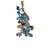
|
|---|---|
| 1 |
|
-Components
-Protein , 12 types, 23 molecules AaCcDdEeFfHhIiKLMNOWXYj
| #1: Protein | Mass: 227854.141 Da / Num. of mol.: 2 / Source method: isolated from a natural source / Source: (natural) #3: Protein | Mass: 41744.512 Da / Num. of mol.: 2 / Source method: isolated from a natural source / Source: (natural) #4: Protein | Mass: 39798.602 Da / Num. of mol.: 2 / Source method: isolated from a natural source / Source: (natural) #5: Protein | Mass: 162658.234 Da / Num. of mol.: 2 / Source method: isolated from a natural source / Source: (natural) #6: Protein | Mass: 36588.625 Da / Num. of mol.: 2 / Source method: isolated from a natural source / Source: (natural) #8: Protein | Mass: 35361.309 Da / Num. of mol.: 2 / Source method: isolated from a natural source / Source: (natural) #9: Protein | Mass: 105398.547 Da / Num. of mol.: 2 / Source method: isolated from a natural source / Source: (natural) #11: Protein | Mass: 322784.344 Da / Num. of mol.: 5 / Source method: isolated from a natural source / Source: (natural) #13: Protein | | Mass: 154922.422 Da / Num. of mol.: 1 / Source method: isolated from a natural source / Source: (natural) #14: Protein | | Mass: 13719.902 Da / Num. of mol.: 1 / Source method: isolated from a natural source Details: The putative protein Nup98 was from the same gene of the chains g/G, whose corresponding peptide is post-translationally cleaved into two separate proteins. Source: (natural) #15: Protein | | Mass: 82570.148 Da / Num. of mol.: 1 / Source method: isolated from a natural source / Source: (natural) #16: Protein | | Mass: 127551.250 Da / Num. of mol.: 1 / Source method: isolated from a natural source / Source: (natural) |
|---|
-Nuclear pore complex protein ... , 4 types, 9 molecules BbGgJPSsp
| #2: Protein | Mass: 75160.047 Da / Num. of mol.: 2 / Source method: isolated from a natural source / Source: (natural) #7: Protein | Mass: 191067.188 Da / Num. of mol.: 2 / Source method: isolated from a natural source / Source: (natural) #10: Protein | | Mass: 111296.109 Da / Num. of mol.: 1 / Source method: isolated from a natural source Details: This entity was identical with chain j. However, N-terminal region cannot be assigned due to disorder. Source: (natural) #12: Protein | Mass: 93565.156 Da / Num. of mol.: 4 / Source method: isolated from a natural source / Source: (natural) |
|---|
-Experimental details
-Experiment
| Experiment | Method: ELECTRON MICROSCOPY |
|---|---|
| EM experiment | Aggregation state: PARTICLE / 3D reconstruction method: single particle reconstruction |
- Sample preparation
| Component | Name: Xenopus laevis Nuclear Pore Complex (NPC) / Type: COMPLEX / Entity ID: #1-#13, #16, #15, #14 / Source: NATURAL |
|---|---|
| Source (natural) | Organism: |
| Buffer solution | pH: 7.5 |
| Specimen | Embedding applied: NO / Shadowing applied: NO / Staining applied: NO / Vitrification applied: YES |
| Vitrification | Cryogen name: ETHANE |
- Electron microscopy imaging
| Experimental equipment |  Model: Titan Krios / Image courtesy: FEI Company |
|---|---|
| Microscopy | Model: FEI TITAN KRIOS |
| Electron gun | Electron source:  FIELD EMISSION GUN / Accelerating voltage: 300 kV / Illumination mode: FLOOD BEAM FIELD EMISSION GUN / Accelerating voltage: 300 kV / Illumination mode: FLOOD BEAM |
| Electron lens | Mode: BRIGHT FIELD / Nominal defocus max: 3000 nm / Nominal defocus min: 1500 nm |
| Image recording | Electron dose: 75 e/Å2 / Film or detector model: GATAN K3 (6k x 4k) |
- Processing
| CTF correction | Type: PHASE FLIPPING AND AMPLITUDE CORRECTION |
|---|---|
| Symmetry | Point symmetry: C1 (asymmetric) |
| 3D reconstruction | Resolution: 3.7 Å / Resolution method: FSC 0.143 CUT-OFF / Num. of particles: 1279270 / Symmetry type: POINT |
| Atomic model building | Space: REAL |
