 Movie
Movie Controller

[English] 日本語
 Yorodumi
- PDB-7ejk: Structure of the alpha2A-adrenergic receptor GoA signaling comple... -
+ Open data

- Basic information
| Entry | Database: PDB / ID: 7ejk | ||||||
|---|---|---|---|---|---|---|---|
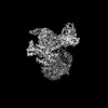
| Title | Structure of the alpha2A-adrenergic receptor GoA signaling complex bound to oxymetazoline | ||||||
 Components Components |
| ||||||
 Keywords Keywords | MEMBRANE PROTEIN / alpha2A-adrenergic receptor / signaling complex / cryo-EM | ||||||
| Function / homology |  Function and homology information Function and homology informationnegative regulation of uterine smooth muscle contraction / alpha2-adrenergic receptor activity / Adrenaline signalling through Alpha-2 adrenergic receptor / alpha-2C adrenergic receptor binding / epinephrine binding / phospholipase C-activating adrenergic receptor signaling pathway / alpha-1B adrenergic receptor binding / negative regulation of norepinephrine secretion / negative regulation of epinephrine secretion / heterotrimeric G-protein binding ...negative regulation of uterine smooth muscle contraction / alpha2-adrenergic receptor activity / Adrenaline signalling through Alpha-2 adrenergic receptor / alpha-2C adrenergic receptor binding / epinephrine binding / phospholipase C-activating adrenergic receptor signaling pathway / alpha-1B adrenergic receptor binding / negative regulation of norepinephrine secretion / negative regulation of epinephrine secretion / heterotrimeric G-protein binding / negative regulation of calcium ion-dependent exocytosis / positive regulation of potassium ion transport / dopaminergic synapse / mu-type opioid receptor binding / thermoception / : / corticotropin-releasing hormone receptor 1 binding / thioesterase binding / fear response / negative regulation of insulin secretion involved in cellular response to glucose stimulus / Surfactant metabolism / adenylate cyclase-inhibiting adrenergic receptor signaling pathway / norepinephrine binding / positive regulation of membrane protein ectodomain proteolysis / Adrenoceptors / intestinal absorption / response to alcohol / G protein-coupled dopamine receptor signaling pathway / adrenergic receptor signaling pathway / positive regulation of wound healing / response to morphine / regulation of heart contraction / negative regulation of calcium ion transport / parallel fiber to Purkinje cell synapse / negative regulation of lipid catabolic process / regulation of vasoconstriction / postsynaptic modulation of chemical synaptic transmission / positive regulation of epidermal growth factor receptor signaling pathway / Rho protein signal transduction / cellular response to hormone stimulus / adenylate cyclase-activating adrenergic receptor signaling pathway / presynaptic active zone membrane / adenylate cyclase-inhibiting serotonin receptor signaling pathway / G protein-coupled serotonin receptor binding / muscle contraction / axon terminus / presynaptic modulation of chemical synaptic transmission / positive regulation of cytokine production / female pregnancy / locomotory behavior / platelet activation / negative regulation of insulin secretion / postsynaptic density membrane / GABA-ergic synapse / vasodilation / epidermal growth factor receptor signaling pathway / G-protein beta/gamma-subunit complex binding / adenylate cyclase-modulating G protein-coupled receptor signaling pathway / adenylate cyclase-inhibiting G protein-coupled receptor signaling pathway / Olfactory Signaling Pathway / Activation of the phototransduction cascade / G protein-coupled acetylcholine receptor signaling pathway / G beta:gamma signalling through PLC beta / Presynaptic function of Kainate receptors / Thromboxane signalling through TP receptor / Activation of G protein gated Potassium channels / Inhibition of voltage gated Ca2+ channels via Gbeta/gamma subunits / G-protein activation / Glucagon signaling in metabolic regulation / Prostacyclin signalling through prostacyclin receptor / G beta:gamma signalling through CDC42 / Synthesis, secretion, and inactivation of Glucagon-like Peptide-1 (GLP-1) / G beta:gamma signalling through BTK / photoreceptor disc membrane / ADP signalling through P2Y purinoceptor 12 / Glucagon-type ligand receptors / Sensory perception of sweet, bitter, and umami (glutamate) taste / Adrenaline,noradrenaline inhibits insulin secretion / Vasopressin regulates renal water homeostasis via Aquaporins / Glucagon-like Peptide-1 (GLP1) regulates insulin secretion / G alpha (z) signalling events / cellular response to catecholamine stimulus / ADP signalling through P2Y purinoceptor 1 / ADORA2B mediated anti-inflammatory cytokines production / G beta:gamma signalling through PI3Kgamma / adenylate cyclase-activating dopamine receptor signaling pathway / Cooperation of PDCL (PhLP1) and TRiC/CCT in G-protein beta folding / glucose homeostasis / GPER1 signaling / cellular response to prostaglandin E stimulus / heterotrimeric G-protein complex / G alpha (12/13) signalling events / G-protein beta-subunit binding / Inactivation, recovery and regulation of the phototransduction cascade / extracellular vesicle / sensory perception of taste / Thrombin signalling through proteinase activated receptors (PARs) / adenylate cyclase-activating G protein-coupled receptor signaling pathway / signaling receptor complex adaptor activity / actin cytoskeleton organization Similarity search - Function | ||||||
| Biological species |  Homo sapiens (human) Homo sapiens (human) | ||||||
| Method | ELECTRON MICROSCOPY / single particle reconstruction / cryo EM / Resolution: 3.4 Å | ||||||
 Authors Authors | Xu, J. / Cao, S. / Liu, Z. / Du, Y. | ||||||
 Citation Citation | Journal: Sci Adv / Year: 2022 Title: Structural insights into ligand recognition, activation, and signaling of the α adrenergic receptor. Authors: Jun Xu / Sheng Cao / Harald Hübner / Dorothée Weikert / Geng Chen / Qiuyuan Lu / Daopeng Yuan / Peter Gmeiner / Zheng Liu / Yang Du /   Abstract: The α adrenergic receptor (αAR) is a G protein (heterotrimeric guanine nucleotide-binding protein)-coupled receptor that mediates important physiological functions in response to the endogenous ...The α adrenergic receptor (αAR) is a G protein (heterotrimeric guanine nucleotide-binding protein)-coupled receptor that mediates important physiological functions in response to the endogenous neurotransmitters norepinephrine and epinephrine, as well as numerous chemically distinct drugs. However, the molecular mechanisms of drug actions remain poorly understood. Here, we report the cryo-electron microscopy structures of the human αAR-GoA complex bound to norepinephrine and three imidazoline derivatives (brimonidine, dexmedetomidine, and oxymetazoline). Together with mutagenesis and functional data, these structures provide important insights into the molecular basis of ligand recognition, activation, and signaling at the αAR. Further structural analyses uncover different molecular determinants between αAR and βARs for recognition of norepinephrine and key regions that determine the G protein coupling selectivity. Overall, our studies provide a framework for understanding the signal transduction of the adrenergic system at the atomic level, which will facilitate rational structure-based discovery of safer and more effective medications for αAR. | ||||||
| History |
|
- Structure visualization
| Structure viewer | Molecule: MolmilJmol/JSmol |
|---|
- Downloads & links
-Download
| PDBx/mmCIF format | 7ejk.cif.gz | 212.6 KB | Display | PDBx/mmCIF format |
|---|---|---|---|---|
| PDB format | pdb7ejk.ent.gz | 161.5 KB | Display | PDB format |
| PDBx/mmJSON format | 7ejk.json.gz | Tree view | PDBx/mmJSON format | |
| Others |  Other downloads Other downloads |
-Validation report
| Arichive directory | https://data.pdbj.org/pub/pdb/validation_reports/ej/7ejkftp://data.pdbj.org/pub/pdb/validation_reports/ej/7ejk | HTTPS FTP |
|---|
-Related structure data
| Related structure data | 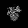 31162MC  7ej0C  7ej8C  7ejaC  7el0 C: citing same article ( M: map data used to model this data |
|---|---|
| Similar structure data |


-Links
 PDBj
PDBj


- Assembly
| Deposited unit | 
|
|---|---|
| 1 |
|
-Components
-Guanine nucleotide-binding protein ... , 3 types, 3 molecules ABG
| #1: Protein | Mass: 40100.500 Da / Num. of mol.: 1 Source method: isolated from a genetically manipulated source Source: (gene. exp.) Homo sapiens (human) / Gene: GNAO1 / Production host:  Trichoplusia ni (cabbage looper) / References: UniProt: P09471 Trichoplusia ni (cabbage looper) / References: UniProt: P09471 |
|---|---|
| #2: Protein | Mass: 38402.867 Da / Num. of mol.: 1 Source method: isolated from a genetically manipulated source Source: (gene. exp.) Homo sapiens (human) / Gene: GNB1 / Production host: Trichoplusia ni (cabbage looper) / References: UniProt: P62873 |
| #3: Protein | Mass: 7861.143 Da / Num. of mol.: 1 Source method: isolated from a genetically manipulated source Source: (gene. exp.) Homo sapiens (human) / Gene: GNG2 / Production host: Trichoplusia ni (cabbage looper) / References: UniProt: P59768 |
-Antibody / Protein / Non-polymers , 3 types, 3 molecules HR
| #4: Antibody | Mass: 32898.781 Da / Num. of mol.: 1 Source method: isolated from a genetically manipulated source Source: (gene. exp.) Trichoplusia ni (cabbage looper) |
|---|---|
| #5: Protein | Mass: 50704.566 Da / Num. of mol.: 1 Source method: isolated from a genetically manipulated source Source: (gene. exp.) Homo sapiens (human) / Gene: ADRA2A, ADRA2R, ADRAR / Production host:  Spodoptera frugiperda (fall armyworm) / References: UniProt: P08913 Spodoptera frugiperda (fall armyworm) / References: UniProt: P08913 |
| #6: Chemical | ChemComp-J5C /  Mass: 260.375 Da / Num. of mol.: 1 / Source method: obtained synthetically / Formula: C16H24N2O / Feature type: SUBJECT OF INVESTIGATION Mass: 260.375 Da / Num. of mol.: 1 / Source method: obtained synthetically / Formula: C16H24N2O / Feature type: SUBJECT OF INVESTIGATION |
-Details
| Has ligand of interest | Y |
|---|---|
| Has protein modification | Y |
-Experimental details
-Experiment
| Experiment | Method: ELECTRON MICROSCOPY |
|---|---|
| EM experiment | Aggregation state: PARTICLE / 3D reconstruction method: single particle reconstruction |
- Sample preparation
| Component |
| ||||||||||||||||||||||||
|---|---|---|---|---|---|---|---|---|---|---|---|---|---|---|---|---|---|---|---|---|---|---|---|---|---|
| Source (natural) |
| ||||||||||||||||||||||||
| Source (recombinant) |
| ||||||||||||||||||||||||
| Buffer solution | pH: 7.5 | ||||||||||||||||||||||||
| Specimen | Embedding applied: YES / Shadowing applied: NO / Staining applied: NO / Vitrification applied: YES | ||||||||||||||||||||||||
| EM embedding | Material: vitreous ice | ||||||||||||||||||||||||
| Vitrification | Cryogen name: ETHANE |
- Electron microscopy imaging
| Experimental equipment |  Model: Titan Krios / Image courtesy: FEI Company |
|---|---|
| Microscopy | Model: FEI TITAN KRIOS |
| Electron gun | Electron source:  FIELD EMISSION GUN / Accelerating voltage: 300 kV / Illumination mode: FLOOD BEAM FIELD EMISSION GUN / Accelerating voltage: 300 kV / Illumination mode: FLOOD BEAM |
| Electron lens | Mode: BRIGHT FIELD / Nominal magnification: 105000 X / Nominal defocus max: 1400 nm / Nominal defocus min: 1000 nm / Calibrated defocus min: 800 nm / Calibrated defocus max: 2000 nm / Cs: 2.7 mm |
| Image recording | Electron dose: 56 e/Å2 / Film or detector model: GATAN K3 BIOQUANTUM (6k x 4k) |
- Processing
| CTF correction | Type: PHASE FLIPPING AND AMPLITUDE CORRECTION |
|---|---|
| 3D reconstruction | Resolution: 3.4 Å / Resolution method: FSC 0.143 CUT-OFF / Num. of particles: 220513 / Symmetry type: POINT |
