 Movie
Movie Controller

[English] 日本語
 Yorodumi
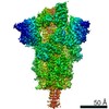
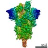
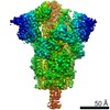
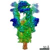
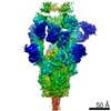
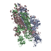
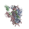
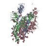
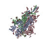
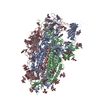
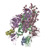
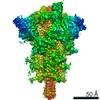
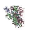
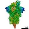
- PDB-7edi: Cryo-EM structure of SARS-CoV-2 S-UK variant (B.1.1.7), two RBD-u... -
+ Open data

- Basic information
| Entry | Database: PDB / ID: 7edi | |||||||||||||||
|---|---|---|---|---|---|---|---|---|---|---|---|---|---|---|---|---|
| Title | Cryo-EM structure of SARS-CoV-2 S-UK variant (B.1.1.7), two RBD-up conformation | |||||||||||||||
 Components Components | Spike glycoprotein | |||||||||||||||
 Keywords Keywords | VIRAL PROTEIN / SARS-CoV-2 / Spike protein | |||||||||||||||
| Function / homology |  Function and homology information Function and homology informationMaturation of spike protein / viral translation / Translation of Structural Proteins / Virion Assembly and Release / host cell surface / host extracellular space / suppression by virus of host tetherin activity / Induction of Cell-Cell Fusion / structural constituent of virion / entry receptor-mediated virion attachment to host cell ...Maturation of spike protein / viral translation / Translation of Structural Proteins / Virion Assembly and Release / host cell surface / host extracellular space / suppression by virus of host tetherin activity / Induction of Cell-Cell Fusion / structural constituent of virion / entry receptor-mediated virion attachment to host cell / host cell endoplasmic reticulum-Golgi intermediate compartment membrane / membrane fusion / receptor-mediated endocytosis of virus by host cell / Attachment and Entry / positive regulation of viral entry into host cell / receptor-mediated virion attachment to host cell / receptor ligand activity / symbiont-mediated suppression of host innate immune response / host cell surface receptor binding / fusion of virus membrane with host plasma membrane / fusion of virus membrane with host endosome membrane / viral envelope / virion attachment to host cell / SARS-CoV-2 activates/modulates innate and adaptive immune responses / host cell plasma membrane / virion membrane / identical protein binding / membrane / plasma membrane Similarity search - Function | |||||||||||||||
| Biological species |   Severe acute respiratory syndrome coronavirus 2 Severe acute respiratory syndrome coronavirus 2 | |||||||||||||||
| Method | ELECTRON MICROSCOPY / single particle reconstruction / cryo EM / Resolution: 3.3 Å | |||||||||||||||
 Authors Authors | Yang, T.J. / Yu, P.Y. / Chang, Y.C. / Wu, H.C. / Hsu, S.T.D. | |||||||||||||||
| Funding support |  Taiwan, 4items Taiwan, 4items
| |||||||||||||||
 Citation Citation | Journal: Nat Struct Mol Biol / Year: 2021 Title: Effect of SARS-CoV-2 B.1.1.7 mutations on spike protein structure and function. Authors: Tzu-Jing Yang / Pei-Yu Yu / Yuan-Chih Chang / Kang-Hao Liang / Hsian-Cheng Tso / Meng-Ru Ho / Wan-Yu Chen / Hsiu-Ting Lin / Han-Chung Wu / Shang-Te Danny Hsu / Abstract: The B.1.1.7 variant of SARS-CoV-2 first detected in the UK harbors amino-acid substitutions and deletions in the spike protein that potentially enhance host angiotensin conversion enzyme 2 (ACE2) ...The B.1.1.7 variant of SARS-CoV-2 first detected in the UK harbors amino-acid substitutions and deletions in the spike protein that potentially enhance host angiotensin conversion enzyme 2 (ACE2) receptor binding and viral immune evasion. Here we report cryo-EM structures of the spike protein of B.1.1.7 in the apo and ACE2-bound forms. The apo form showed one or two receptor-binding domains (RBDs) in the open conformation, without populating the fully closed state. All three RBDs were engaged in ACE2 binding. The B.1.1.7-specific A570D mutation introduces a molecular switch that could modulate the opening and closing of the RBD. The N501Y mutation introduces a π-π interaction that enhances RBD binding to ACE2 and abolishes binding of a potent neutralizing antibody (nAb). Cryo-EM also revealed how a cocktail of two nAbs simultaneously bind to all three RBDs, and demonstrated the potency of the nAb cocktail to neutralize different SARS-CoV-2 pseudovirus strains, including B.1.1.7. | |||||||||||||||
| History |
|
- Structure visualization
| Movie |
Movie viewer |
|---|---|
| Structure viewer | Molecule: MolmilJmol/JSmol |
- Downloads & links
-Download
| PDBx/mmCIF format | 7edi.cif.gz | 564.8 KB | Display | PDBx/mmCIF format |
|---|---|---|---|---|
| PDB format | pdb7edi.ent.gz | 469.3 KB | Display | PDB format |
| PDBx/mmJSON format | 7edi.json.gz | Tree view | PDBx/mmJSON format | |
| Others |  Other downloads Other downloads |
-Validation report
| Summary document | 7edi_validation.pdf.gz | 2.3 MB | Display | wwPDB validaton report |
|---|---|---|---|---|
| Full document | 7edi_full_validation.pdf.gz | 2.3 MB | Display | |
| Data in XML | 7edi_validation.xml.gz | 75.7 KB | Display | |
| Data in CIF | 7edi_validation.cif.gz | 118.2 KB | Display | |
| Arichive directory | https://data.pdbj.org/pub/pdb/validation_reports/ed/7ediftp://data.pdbj.org/pub/pdb/validation_reports/ed/7edi | HTTPS FTP |
-Related structure data
| Related structure data | 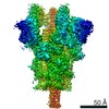 31072MC  7edfC  7edgC  7edhC 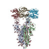 7edjC 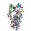 7eh5C C: citing same article ( M: map data used to model this data |
|---|---|
| Similar structure data |
-Links
 PDBj
PDBj


- Assembly
| Deposited unit | 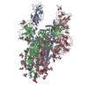
|
|---|---|
| 1 |
|
-Components
| #1: Protein | Mass: 142662.797 Da / Num. of mol.: 3 Source method: isolated from a genetically manipulated source Source: (gene. exp.) Severe acute respiratory syndrome coronavirus 2Gene: S, 2 / Production host:  Homo sapiens (human) / References: UniProt: P0DTC2 Homo sapiens (human) / References: UniProt: P0DTC2#2: Polysaccharide | 2-acetamido-2-deoxy-beta-D-glucopyranose-(1-4)-2-acetamido-2-deoxy-beta-D-glucopyranose Source method: isolated from a genetically manipulated source #3: Sugar | ChemComp-NAG /   Type: D-saccharide, beta linking / Mass: 221.208 Da / Num. of mol.: 16 / Source method: obtained synthetically / Formula: C8H15NO6 Type: D-saccharide, beta linking / Mass: 221.208 Da / Num. of mol.: 16 / Source method: obtained synthetically / Formula: C8H15NO6Has ligand of interest | N | Sequence details | The two deletions, including residues 69-70 and residue 144 are designed for this study. The ...The two deletions, including residues 69-70 and residue 144 are designed for this study. The residues, including Y498, D567, G611, H678, I713, A979 and H1115, are designed for this study. The residues 679-682 (RAAA) and residues 983-984 (PP) are designed for protein stabilization. | |
|---|
-Experimental details
-Experiment
| Experiment | Method: ELECTRON MICROSCOPY |
|---|---|
| EM experiment | Aggregation state: PARTICLE / 3D reconstruction method: single particle reconstruction |
- Sample preparation
| Component | Name: SARS-CoV-2 spike glycoprotein / Type: ORGANELLE OR CELLULAR COMPONENT / Details: UK variant (B.1.1.7), two RBP-up conformation / Entity ID: #1 / Source: RECOMBINANT | ||||||||||||||||||||
|---|---|---|---|---|---|---|---|---|---|---|---|---|---|---|---|---|---|---|---|---|---|
| Molecular weight | Value: 0.54 MDa / Experimental value: YES | ||||||||||||||||||||
| Source (natural) | Organism: Severe acute respiratory syndrome coronavirus 2 | ||||||||||||||||||||
| Source (recombinant) | Organism: Homo sapiens (human) | ||||||||||||||||||||
| Buffer solution | pH: 7.6 | ||||||||||||||||||||
| Buffer component |
| ||||||||||||||||||||
| Specimen | Conc.: 1 mg/ml / Embedding applied: NO / Shadowing applied: NO / Staining applied: NO / Vitrification applied: YES | ||||||||||||||||||||
| Vitrification | Instrument: FEI VITROBOT MARK IV / Cryogen name: ETHANE / Humidity: 100 % / Chamber temperature: 277 K Details: blot for 2.5 seconds before plunging; blot force: 0; waiting time: 30s |
- Electron microscopy imaging
| Experimental equipment |  Model: Titan Krios / Image courtesy: FEI Company |
|---|---|
| Microscopy | Model: FEI TITAN KRIOS |
| Electron gun | Electron source:  FIELD EMISSION GUN / Accelerating voltage: 300 kV / Illumination mode: FLOOD BEAM FIELD EMISSION GUN / Accelerating voltage: 300 kV / Illumination mode: FLOOD BEAM |
| Electron lens | Mode: BRIGHT FIELD / Cs: 2.7 mm |
| Specimen holder | Cryogen: NITROGEN |
| Image recording | Electron dose: 1 e/Å2 / Film or detector model: GATAN K3 (6k x 4k) |
- Processing
| Software | Name: PHENIX / Version: 1.19.1_4122: / Classification: refinement | ||||||||||||||||||||||||
|---|---|---|---|---|---|---|---|---|---|---|---|---|---|---|---|---|---|---|---|---|---|---|---|---|---|
| EM software |
| ||||||||||||||||||||||||
| CTF correction | Type: PHASE FLIPPING AND AMPLITUDE CORRECTION | ||||||||||||||||||||||||
| Symmetry | Point symmetry: C1 (asymmetric) | ||||||||||||||||||||||||
| 3D reconstruction | Resolution: 3.3 Å / Resolution method: FSC 0.143 CUT-OFF / Num. of particles: 158098 / Symmetry type: POINT | ||||||||||||||||||||||||
| Refine LS restraints |
|
