 Movie
Movie Controller

[English] 日本語
 Yorodumi
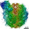
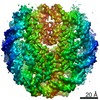
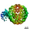
- PDB-7ea5: Yeast Set2 bound to a nucleosome containing oncohistone mutations -
+ Open data

- Basic information
| Entry | Database: PDB / ID: 7ea5 | |||||||||||||||
|---|---|---|---|---|---|---|---|---|---|---|---|---|---|---|---|---|
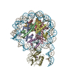
| Title | Yeast Set2 bound to a nucleosome containing oncohistone mutations | |||||||||||||||
 Components Components |
| |||||||||||||||
 Keywords Keywords | GENE REGULATION / methyltransferase / Set2 / nucleosome / H3K36M mutation | |||||||||||||||
| Function / homology |  Function and homology information Function and homology information[histone H3]-lysine36 N-trimethyltransferase / histone H3K36 methyltransferase activity / structural constituent of chromatin / nucleosome / heterochromatin formation / chromosome / protein heterodimerization activity / regulation of DNA-templated transcription / DNA binding / nucleus Similarity search - Function | |||||||||||||||
| Biological species |  synthetic construct (others) | |||||||||||||||
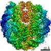
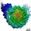
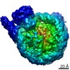
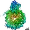
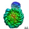
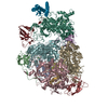
| Method | ELECTRON MICROSCOPY / single particle reconstruction / cryo EM / Resolution: 3.3 Å | |||||||||||||||
 Authors Authors | Jing, H. / Liu, Y. | |||||||||||||||
| Funding support |  China, 3items China, 3items
| |||||||||||||||
 Citation Citation | Journal: Cell Discov / Year: 2021 Title: Cryo-EM structure of SETD2/Set2 methyltransferase bound to a nucleosome containing oncohistone mutations. Authors: Yingying Liu / Yanjun Zhang / Han Xue / Mi Cao / Guohui Bai / Zongkai Mu / Yanli Yao / Shuyang Sun / Dong Fang / Jing Huang / Abstract: Substitution of lysine 36 with methionine in histone H3.3 (H3.3K36M) is an oncogenic mutation that inhibits SETD2-mediated histone H3K36 tri-methylation in tumors. To investigate how the oncohistone ...Substitution of lysine 36 with methionine in histone H3.3 (H3.3K36M) is an oncogenic mutation that inhibits SETD2-mediated histone H3K36 tri-methylation in tumors. To investigate how the oncohistone mutation affects the function of SETD2 at the nucleosome level, we determined the cryo-EM structure of human SETD2 associated with an H3.3K36M nucleosome and cofactor S-adenosylmethionine (SAM), and revealed that SETD2 is attached to the N-terminal region of histone H3 and the nucleosome DNA at superhelix location 1, accompanied with the partial unwrapping of nucleosome DNA to expose the SETD2-binding site. These structural features were also observed in the previous cryo-EM structure of the fungal Set2-nucleosome complex. By contrast with the stable association of SETD2 with the H3.3K36M nucleosome, the EM densities of SETD2 could not be observed on the wild-type nucleosome surface, suggesting that the association of SETD2 with wild-type nucleosome might be transient. The linker histone H1, which stabilizes the wrapping of nucleosome DNA at the entry/exit sites, exhibits an inhibitory effect on the activities of SETD2 and displays inversely correlated genome distributions with that of the H3K36me3 marks. Cryo-EM analysis of yeast H3K36 methyltransferase Set2 complexed with nucleosomes further revealed evolutionarily conserved structural features for nucleosome recognition in eukaryotes, and provides insights into the mechanism of activity regulation. These findings have advanced our understanding of the structural basis for the tumorigenesis mechanism of the H3.3K36M mutation and highlight the effect of nucleosome conformation on the regulation of histone modification. | |||||||||||||||
| History |
|
- Structure visualization
| Movie |
Movie viewer |
|---|---|
| Structure viewer | Molecule: MolmilJmol/JSmol |
- Downloads & links
-Download
| PDBx/mmCIF format | 7ea5.cif.gz | 326.8 KB | Display | PDBx/mmCIF format |
|---|---|---|---|---|
| PDB format | pdb7ea5.ent.gz | 241.3 KB | Display | PDB format |
| PDBx/mmJSON format | 7ea5.json.gz | Tree view | PDBx/mmJSON format | |
| Others |  Other downloads Other downloads |
-Validation report
| Arichive directory | https://data.pdbj.org/pub/pdb/validation_reports/ea/7ea5ftp://data.pdbj.org/pub/pdb/validation_reports/ea/7ea5 | HTTPS FTP |
|---|
-Related structure data
| Related structure data | 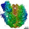 31039MC 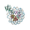 7ea8C M: map data used to model this data C: citing same article ( |
|---|---|
| Similar structure data |
-Links
 PDBj
PDBj


- Assembly
| Deposited unit | 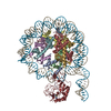
|
|---|---|
| 1 |
|
-Components
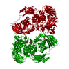
-Protein , 5 types, 9 molecules AEBFCGDHK
| #1: Protein | Mass: 11847.916 Da / Num. of mol.: 2 / Mutation: K37M Source method: isolated from a genetically manipulated source Source: (gene. exp.)  #2: Protein | Mass: 8853.342 Da / Num. of mol.: 2 Source method: isolated from a genetically manipulated source Source: (gene. exp.) #3: Protein | Mass: 11494.393 Da / Num. of mol.: 2 Source method: isolated from a genetically manipulated source Source: (gene. exp.) Gene: h2ac14.L, h2ac14, hist1h2aj, LOC494591, XELAEV_18003602mg Production host: #4: Protein | Mass: 10406.954 Da / Num. of mol.: 2 Source method: isolated from a genetically manipulated source Source: (gene. exp.) #7: Protein | | Mass: 26699.021 Da / Num. of mol.: 1 Source method: isolated from a genetically manipulated source Source: (gene. exp.) Gene: SCNYR20_0009003900, SCP684_0009003900 / Production host: References: UniProt: A0A6V8RR65, [histone H3]-lysine36 N-trimethyltransferase |
|---|
-DNA chain , 2 types, 2 molecules IJ
| #5: DNA chain | Mass: 44521.367 Da / Num. of mol.: 1 / Source method: obtained synthetically / Source: (synth.) synthetic construct (others) |
|---|---|
| #6: DNA chain | Mass: 44992.648 Da / Num. of mol.: 1 / Source method: obtained synthetically / Source: (synth.) synthetic construct (others) |
-Non-polymers , 2 types, 4 molecules 

| #8: Chemical |  Mass: 65.409 Da / Num. of mol.: 3 / Source method: obtained synthetically / Formula: Zn Mass: 65.409 Da / Num. of mol.: 3 / Source method: obtained synthetically / Formula: Zn#9: Chemical | ChemComp-SAM / |  Mass: 398.437 Da / Num. of mol.: 1 / Source method: obtained synthetically / Formula: C15H22N6O5S Mass: 398.437 Da / Num. of mol.: 1 / Source method: obtained synthetically / Formula: C15H22N6O5S |
|---|
-Details
| Has ligand of interest | N |
|---|---|
| Has protein modification | N |
-Experimental details
-Experiment
| Experiment | Method: ELECTRON MICROSCOPY |
|---|---|
| EM experiment | Aggregation state: PARTICLE / 3D reconstruction method: single particle reconstruction |
- Sample preparation
| Component | Name: The Set2-nucleosome(H3K36M)complex structure / Type: COMPLEX / Entity ID: #1-#7 / Source: MULTIPLE SOURCES |
|---|---|
| Molecular weight | Experimental value: NO |
| Source (natural) | Organism: |
| Source (recombinant) | Organism: |
| Buffer solution | pH: 7.5 |
| Specimen | Conc.: 0.3 mg/ml / Embedding applied: NO / Shadowing applied: NO / Staining applied: NO / Vitrification applied: YES |
| Vitrification | Cryogen name: ETHANE |
- Electron microscopy imaging
| Experimental equipment |  Model: Titan Krios / Image courtesy: FEI Company |
|---|---|
| Microscopy | Model: FEI TITAN KRIOS |
| Electron gun | Electron source:  FIELD EMISSION GUN / Accelerating voltage: 300 kV / Illumination mode: FLOOD BEAM FIELD EMISSION GUN / Accelerating voltage: 300 kV / Illumination mode: FLOOD BEAM |
| Electron lens | Mode: BRIGHT FIELD |
| Image recording | Electron dose: 1.5625 e/Å2 / Film or detector model: GATAN K3 (6k x 4k) |
- Processing
| Software |
| ||||||||||||||||||||||||
|---|---|---|---|---|---|---|---|---|---|---|---|---|---|---|---|---|---|---|---|---|---|---|---|---|---|
| CTF correction | Type: PHASE FLIPPING AND AMPLITUDE CORRECTION | ||||||||||||||||||||||||
| 3D reconstruction | Resolution: 3.3 Å / Resolution method: FSC 0.143 CUT-OFF / Num. of particles: 225474 / Symmetry type: POINT | ||||||||||||||||||||||||
| Refinement | Stereochemistry target values: GeoStd + Monomer Library | ||||||||||||||||||||||||
| Refine LS restraints |
|
