 Movie
Movie Controller


+ Open data

- Basic information
| Entry | Database: PDB / ID: 7dxi | ||||||
|---|---|---|---|---|---|---|---|
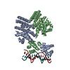
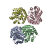
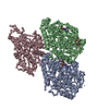
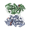
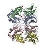
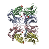
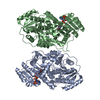
| Title | Structure of Drosophila melanogaster GlcNAc-1-phosphotransferase | ||||||
 Components Components | FI02838p | ||||||
 Keywords Keywords | TRANSFERASE / GNPTAB / GlcNAc-1-phosphotransferase / SUGAR BINDING PROTEIN | ||||||
| Function / homology |  Function and homology information Function and homology informationN-glycan processing to lysosome / UDP-N-acetylglucosamine-lysosomal-enzyme N-acetylglucosaminephosphotransferase activity / carbohydrate phosphorylation / membrane => GO:0016020 / Golgi apparatus Similarity search - Function | ||||||
| Biological species |  | ||||||
| Method | ELECTRON MICROSCOPY / single particle reconstruction / cryo EM / Resolution: 3.53 Å | ||||||
 Authors Authors | Du, S. / Xiao, J. / Guopeng, W. | ||||||
 Citation Citation | Journal: J Biol Chem / Year: 2022 Title: Structural insights into how GlcNAc-1-phosphotransferase directs lysosomal protein transport. Authors: Shuo Du / Guopeng Wang / Zhiying Zhang / Chengying Ma / Ning Gao / Junyu Xiao /  Abstract: GlcNAc-1-phosphotransferase catalyzes the initial step in the formation of the mannose-6-phosphate tag that labels ∼60 lysosomal proteins for transport. Mutations in GlcNAc-1-phosphotransferase are ...GlcNAc-1-phosphotransferase catalyzes the initial step in the formation of the mannose-6-phosphate tag that labels ∼60 lysosomal proteins for transport. Mutations in GlcNAc-1-phosphotransferase are known to cause lysosomal storage disorders such as mucolipidoses. However, the molecular mechanism of GlcNAc-1-phosphotransferase activity remains unclear. Mammalian GlcNAc-1-phosphotransferases are α2β2γ2 hexamers in which the core catalytic α- and β-subunits are derived from the GNPTAB (N-acetylglucosamine-1-phosphate transferase subunits alpha and beta) gene. Here, we present the cryo-electron microscopy structure of the Drosophila melanogaster GNPTAB homolog, DmGNPTAB. We identified four conserved regions located far apart in the sequence that fold into the catalytic domain, which exhibits structural similarity to that of the UDP-glucose glycoprotein glucosyltransferase. Comparison with UDP-glucose glycoprotein glucosyltransferase also revealed a putative donor substrate-binding site, and the functional requirements of critical residues in human GNPTAB were validated using GNPTAB-knockout cells. Finally, we show that DmGNPTAB forms a homodimer that is evolutionarily conserved and that perturbing the dimer interface undermines the maturation and activity of human GNPTAB. These results provide important insights into GlcNAc-1-phosphotransferase function and related diseases. | ||||||
| History |
|
- Structure visualization
| Movie |
Movie viewer |
|---|---|
| Structure viewer | Molecule: MolmilJmol/JSmol |
- Downloads & links
-Download
| PDBx/mmCIF format | 7dxi.cif.gz | 147.9 KB | Display | PDBx/mmCIF format |
|---|---|---|---|---|
| PDB format | pdb7dxi.ent.gz | 115.1 KB | Display | PDB format |
| PDBx/mmJSON format | 7dxi.json.gz | Tree view | PDBx/mmJSON format | |
| Others |  Other downloads Other downloads |
-Validation report
| Summary document | 7dxi_validation.pdf.gz | 676.6 KB | Display | wwPDB validaton report |
|---|---|---|---|---|
| Full document | 7dxi_full_validation.pdf.gz | 682.4 KB | Display | |
| Data in XML | 7dxi_validation.xml.gz | 27.2 KB | Display | |
| Data in CIF | 7dxi_validation.cif.gz | 37.3 KB | Display | |
| Arichive directory | https://data.pdbj.org/pub/pdb/validation_reports/dx/7dxiftp://data.pdbj.org/pub/pdb/validation_reports/dx/7dxi | HTTPS FTP |
-Related structure data
| Related structure data | 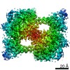 30910MC M: map data used to model this data C: citing same article ( |
|---|---|
| Similar structure data |


-Links
 PDBj
PDBj- Assembly
| Deposited unit | 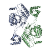
|
|---|---|
| 1 |
|
-Components
| #1: Protein | Mass: 67925.500 Da / Num. of mol.: 2 Source method: isolated from a genetically manipulated source Source: (gene. exp.) Trichoplusia ni (cabbage looper) / References: UniProt: A1Z7S7#2: Chemical |   Mass: 40.078 Da / Num. of mol.: 2 / Source method: obtained synthetically / Formula: Ca / Feature type: SUBJECT OF INVESTIGATION Mass: 40.078 Da / Num. of mol.: 2 / Source method: obtained synthetically / Formula: Ca / Feature type: SUBJECT OF INVESTIGATIONHas ligand of interest | Y | |
|---|
-Experimental details
-Experiment
| Experiment | Method: ELECTRON MICROSCOPY |
|---|---|
| EM experiment | Aggregation state: PARTICLE / 3D reconstruction method: single particle reconstruction |
- Sample preparation
| Component | Name: gnpt / Type: COMPLEX / Entity ID: #1 / Source: RECOMBINANT |
|---|---|
| Source (natural) | Organism: |
| Source (recombinant) | Organism: Trichoplusia ni (cabbage looper) |
| Buffer solution | pH: 8 |
| Specimen | Embedding applied: NO / Shadowing applied: NO / Staining applied: NO / Vitrification applied: YES |
| Vitrification | Cryogen name: ETHANE |
- Electron microscopy imaging
| Experimental equipment |  Model: Titan Krios / Image courtesy: FEI Company |
|---|---|
| Microscopy | Model: FEI TITAN KRIOS |
| Electron gun | Electron source:  FIELD EMISSION GUN / Accelerating voltage: 300 kV / Illumination mode: FLOOD BEAM FIELD EMISSION GUN / Accelerating voltage: 300 kV / Illumination mode: FLOOD BEAM |
| Electron lens | Mode: BRIGHT FIELD |
| Image recording | Average exposure time: 3.2 sec. / Electron dose: 60.29 e/Å2 / Film or detector model: GATAN K2 QUANTUM (4k x 4k) |
- Processing
| Software | Name: PHENIX / Version: 1.18.2_3874: / Classification: refinement | ||||||||||||||||||||||||
|---|---|---|---|---|---|---|---|---|---|---|---|---|---|---|---|---|---|---|---|---|---|---|---|---|---|
| CTF correction | Type: PHASE FLIPPING AND AMPLITUDE CORRECTION | ||||||||||||||||||||||||
| 3D reconstruction | Resolution: 3.53 Å / Resolution method: FSC 0.143 CUT-OFF / Num. of particles: 131301 / Symmetry type: POINT | ||||||||||||||||||||||||
| Refine LS restraints |
|
