 Movie
Movie Controller

[English] 日本語
 Yorodumi
- PDB-7cro: NSD2 bearing E1099K/T1150A dual mutation in complex with 187-bp NCP -
+ Open data

- Basic information
| Entry | Database: PDB / ID: 7cro | ||||||||||||||||||||||||||||||||||||||||||||||||
|---|---|---|---|---|---|---|---|---|---|---|---|---|---|---|---|---|---|---|---|---|---|---|---|---|---|---|---|---|---|---|---|---|---|---|---|---|---|---|---|---|---|---|---|---|---|---|---|---|---|
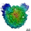
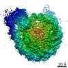
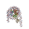
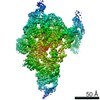
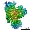
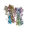
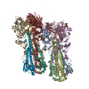
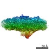
| Title | NSD2 bearing E1099K/T1150A dual mutation in complex with 187-bp NCP | ||||||||||||||||||||||||||||||||||||||||||||||||
 Components Components |
| ||||||||||||||||||||||||||||||||||||||||||||||||
 Keywords Keywords | GENE REGULATION / nucleosome complex / histone methyltransferase / DNA | ||||||||||||||||||||||||||||||||||||||||||||||||
| Function / homology |  Function and homology information Function and homology informationatrial septum secundum morphogenesis / [histone H3]-lysine36 N-dimethyltransferase / histone H4K20 methyltransferase activity / regulation of double-strand break repair via nonhomologous end joining / histone H3K36 dimethyltransferase activity / histone H3K36 trimethyltransferase activity / positive regulation of isotype switching to IgA isotypes / atrial septum primum morphogenesis / membranous septum morphogenesis / regulation of establishment of protein localization ...atrial septum secundum morphogenesis / [histone H3]-lysine36 N-dimethyltransferase / histone H4K20 methyltransferase activity / regulation of double-strand break repair via nonhomologous end joining / histone H3K36 dimethyltransferase activity / histone H3K36 trimethyltransferase activity / positive regulation of isotype switching to IgA isotypes / atrial septum primum morphogenesis / membranous septum morphogenesis / regulation of establishment of protein localization / histone H3K36 methyltransferase activity / histone H3 methyltransferase activity / Nonhomologous End-Joining (NHEJ) / G2/M DNA damage checkpoint / bone development / PKMTs methylate histone lysines / structural constituent of chromatin / nucleosome / double-strand break repair / heterochromatin formation / nucleosome assembly / Recruitment and ATM-mediated phosphorylation of repair and signaling proteins at DNA double strand breaks / Processing of DNA double-strand break ends / methylation / sequence-specific DNA binding / protein heterodimerization activity / chromatin binding / regulation of DNA-templated transcription / chromatin / nucleolus / negative regulation of transcription by RNA polymerase II / DNA binding / zinc ion binding / nucleoplasm / nucleus / cytoplasm Similarity search - Function | ||||||||||||||||||||||||||||||||||||||||||||||||
| Biological species |  Homo sapiens (human) Homo sapiens (human) | ||||||||||||||||||||||||||||||||||||||||||||||||
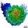
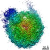
| Method | ELECTRON MICROSCOPY / single particle reconstruction / cryo EM / Resolution: 3.75 Å | ||||||||||||||||||||||||||||||||||||||||||||||||
 Authors Authors | Li, W. / Tian, W. / Yuan, G. / Deng, P. / Gozani, O. / Patel, D. / Wang, Z. | ||||||||||||||||||||||||||||||||||||||||||||||||
| Funding support |  China, China,  United States, 6items United States, 6items
| ||||||||||||||||||||||||||||||||||||||||||||||||
 Citation Citation | Journal: Nature / Year: 2021 Title: Molecular basis of nucleosomal H3K36 methylation by NSD methyltransferases. Authors: Wanqiu Li / Wei Tian / Gang Yuan / Pujuan Deng / Deepanwita Sengupta / Zhongjun Cheng / Yinghua Cao / Jiahao Ren / Yan Qin / Yuqiao Zhou / Yulin Jia / Or Gozani / Dinshaw J Patel / Zhanxin Wang / Abstract: Histone methyltransferases of the nuclear receptor-binding SET domain protein (NSD) family, including NSD1, NSD2 and NSD3, have crucial roles in chromatin regulation and are implicated in oncogenesis. ...Histone methyltransferases of the nuclear receptor-binding SET domain protein (NSD) family, including NSD1, NSD2 and NSD3, have crucial roles in chromatin regulation and are implicated in oncogenesis. NSD enzymes exhibit an autoinhibitory state that is relieved by binding to nucleosomes, enabling dimethylation of histone H3 at Lys36 (H3K36). However, the molecular basis that underlies this mechanism is largely unknown. Here we solve the cryo-electron microscopy structures of NSD2 and NSD3 bound to mononucleosomes. We find that binding of NSD2 and NSD3 to mononucleosomes causes DNA near the linker region to unwrap, which facilitates insertion of the catalytic core between the histone octamer and the unwrapped segment of DNA. A network of DNA- and histone-specific contacts between NSD2 or NSD3 and the nucleosome precisely defines the position of the enzyme on the nucleosome, explaining the specificity of methylation to H3K36. Intermolecular contacts between NSD proteins and nucleosomes are altered by several recurrent cancer-associated mutations in NSD2 and NSD3. NSDs that contain these mutations are catalytically hyperactive in vitro and in cells, and their ectopic expression promotes the proliferation of cancer cells and the growth of xenograft tumours. Together, our research provides molecular insights into the nucleosome-based recognition and histone-modification mechanisms of NSD2 and NSD3, which could lead to strategies for therapeutic targeting of proteins of the NSD family. | ||||||||||||||||||||||||||||||||||||||||||||||||
| History |
|
- Structure visualization
| Movie |
Movie viewer |
|---|---|
| Structure viewer | Molecule: MolmilJmol/JSmol |
- Downloads & links
-Download
| PDBx/mmCIF format | 7cro.cif.gz | 455.4 KB | Display | PDBx/mmCIF format |
|---|---|---|---|---|
| PDB format | pdb7cro.ent.gz | 327 KB | Display | PDB format |
| PDBx/mmJSON format | 7cro.json.gz | Tree view | PDBx/mmJSON format | |
| Others |  Other downloads Other downloads |
-Validation report
| Arichive directory | https://data.pdbj.org/pub/pdb/validation_reports/cr/7croftp://data.pdbj.org/pub/pdb/validation_reports/cr/7cro | HTTPS FTP |
|---|
-Related structure data
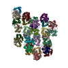
| Related structure data |  30453MC 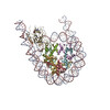 7crpC 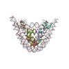 7crqC  7crrC M: map data used to model this data C: citing same article ( |
|---|---|
| Similar structure data |

-Links
 PDBj
PDBj


- Assembly
| Deposited unit | 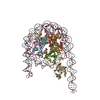
|
|---|---|
| 1 |
|
-Components
-Protein , 5 types, 9 molecules MEBFCGDHI
| #1: Protein | Mass: 15223.779 Da / Num. of mol.: 2 / Mutation: K36Nle, M90Nle, M120Nle Source method: isolated from a genetically manipulated source Source: (gene. exp.)  #2: Protein | Mass: 11263.231 Da / Num. of mol.: 2 Source method: isolated from a genetically manipulated source Source: (gene. exp.) #3: Protein | Mass: 13978.241 Da / Num. of mol.: 2 Source method: isolated from a genetically manipulated source Source: (gene. exp.) #4: Protein | Mass: 13524.752 Da / Num. of mol.: 2 Source method: isolated from a genetically manipulated source Source: (gene. exp.) Gene: h2bc12, hist1h2bk / Production host: #7: Protein | | Mass: 79688.844 Da / Num. of mol.: 1 / Mutation: E1099K, T1150A Source method: isolated from a genetically manipulated source Source: (gene. exp.) Homo sapiens (human) / Gene: NSD2, KIAA1090, MMSET, TRX5, WHSC1 / Production host:  Baculovirus transfer vector pFASTBAC1 Baculovirus transfer vector pFASTBAC1References: UniProt: O96028, [histone H3]-lysine36 N-dimethyltransferase |
|---|
-DNA chain , 2 types, 2 molecules AK
| #5: DNA chain | Mass: 57488.551 Da / Num. of mol.: 1 Source method: isolated from a genetically manipulated source Source: (gene. exp.) |
|---|---|
| #6: DNA chain | Mass: 57982.918 Da / Num. of mol.: 1 Source method: isolated from a genetically manipulated source Source: (gene. exp.) |
-Non-polymers , 2 types, 4 molecules 

| #8: Chemical | ChemComp-SAM /  Mass: 398.437 Da / Num. of mol.: 1 / Source method: obtained synthetically / Formula: C15H22N6O5S / Feature type: SUBJECT OF INVESTIGATION Mass: 398.437 Da / Num. of mol.: 1 / Source method: obtained synthetically / Formula: C15H22N6O5S / Feature type: SUBJECT OF INVESTIGATION |
|---|---|
| #9: Chemical |  Mass: 65.409 Da / Num. of mol.: 3 / Source method: obtained synthetically / Formula: Zn / Feature type: SUBJECT OF INVESTIGATION Mass: 65.409 Da / Num. of mol.: 3 / Source method: obtained synthetically / Formula: Zn / Feature type: SUBJECT OF INVESTIGATION |
-Details
| Has ligand of interest | Y |
|---|---|
| Has protein modification | Y |
-Experimental details
-Experiment
| Experiment | Method: ELECTRON MICROSCOPY |
|---|---|
| EM experiment | Aggregation state: PARTICLE / 3D reconstruction method: single particle reconstruction |
- Sample preparation
| Component |
| ||||||||||||||||||||||||||||||
|---|---|---|---|---|---|---|---|---|---|---|---|---|---|---|---|---|---|---|---|---|---|---|---|---|---|---|---|---|---|---|---|
| Molecular weight | Value: 0.3 MDa / Experimental value: YES | ||||||||||||||||||||||||||||||
| Source (natural) |
| ||||||||||||||||||||||||||||||
| Source (recombinant) |
| ||||||||||||||||||||||||||||||
| Buffer solution | pH: 7.5 | ||||||||||||||||||||||||||||||
| Specimen | Conc.: 0.3 mg/ml / Embedding applied: NO / Shadowing applied: NO / Staining applied: NO / Vitrification applied: YES / Details: The sample was monodisperse. | ||||||||||||||||||||||||||||||
| Vitrification | Instrument: FEI VITROBOT MARK IV / Cryogen name: ETHANE / Humidity: 100 % / Chamber temperature: 281 K Details: blotted for 3 s before being plunged into liquid ethane |
- Electron microscopy imaging
| Experimental equipment |  Model: Titan Krios / Image courtesy: FEI Company |
|---|---|
| Microscopy | Model: FEI TITAN KRIOS |
| Electron gun | Electron source:  FIELD EMISSION GUN / Accelerating voltage: 300 kV / Illumination mode: FLOOD BEAM FIELD EMISSION GUN / Accelerating voltage: 300 kV / Illumination mode: FLOOD BEAM |
| Electron lens | Mode: BRIGHT FIELD / Cs: 2.7 mm / C2 aperture diameter: 70 µm |
| Specimen holder | Cryogen: NITROGEN / Specimen holder model: FEI TITAN KRIOS AUTOGRID HOLDER |
| Image recording | Electron dose: 50 e/Å2 / Detector mode: SUPER-RESOLUTION / Film or detector model: GATAN K2 SUMMIT (4k x 4k) |
| EM imaging optics | Energyfilter name: GIF Bioquantum / Energyfilter slit width: 20 eV |
| Image scans | Movie frames/image: 32 |
- Processing
| Software | Name: PHENIX / Version: 1.16_3549: / Classification: refinement | ||||||||||||||||||||||||
|---|---|---|---|---|---|---|---|---|---|---|---|---|---|---|---|---|---|---|---|---|---|---|---|---|---|
| EM software | Name: PHENIX / Category: model refinement | ||||||||||||||||||||||||
| CTF correction | Type: PHASE FLIPPING AND AMPLITUDE CORRECTION | ||||||||||||||||||||||||
| 3D reconstruction | Resolution: 3.75 Å / Resolution method: FSC 0.143 CUT-OFF / Num. of particles: 71116 / Symmetry type: POINT | ||||||||||||||||||||||||
| Refine LS restraints |
|
