 Movie
Movie Controller

[English] 日本語
 Yorodumi
- PDB-7aoy: Helical arrangement of Bunyamwera virus nucleocapsid protein with... -
+ Open data

- Basic information
| Entry | Database: PDB / ID: 7aoy | ||||||
|---|---|---|---|---|---|---|---|
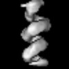
| Title | Helical arrangement of Bunyamwera virus nucleocapsid protein within a native ribonucleoprotein | ||||||
 Components Components | Nucleoprotein | ||||||
 Keywords Keywords | VIRAL PROTEIN / helix / ribonucleoprotein / nucleocapsid / virus | ||||||
| Function / homology |  Function and homology information Function and homology informationhelical viral capsid / symbiont-mediated suppression of host gene expression / viral nucleocapsid / ribonucleoprotein complex / RNA binding Similarity search - Function | ||||||
| Biological species |  Bunyamwera virus Bunyamwera virus | ||||||
| Method | ELECTRON MICROSCOPY / single particle reconstruction / cryo EM / Resolution: 13 Å | ||||||
 Authors Authors | Hopkins, F.R. / Fontana, J. / Barr, J.N. | ||||||
| Funding support |  United Kingdom, 1items United Kingdom, 1items
| ||||||
 Citation Citation | Journal: mBio / Year: 2022 Title: The Native Orthobunyavirus Ribonucleoprotein Possesses a Helical Architecture. Authors: Francis R Hopkins / Beatriz Álvarez-Rodríguez / George R Heath / Kyriakoulla Panayi / Samantha Hover / Thomas A Edwards / John N Barr / Juan Fontana / Abstract: The order is the largest group of negative-sense RNA viruses, containing many lethal human pathogens for which approved anti-infective measures are not available. The bunyavirus genome consists of ...The order is the largest group of negative-sense RNA viruses, containing many lethal human pathogens for which approved anti-infective measures are not available. The bunyavirus genome consists of multiple negative-sense RNA segments enwrapped by the virus-encoded nucleocapsid protein (NP), which together with the viral polymerase form ribonucleoproteins (RNPs). RNPs represent substrates for RNA synthesis and virion assembly, which require inherent flexibility, consistent with the appearance of RNPs spilled from virions. These observations have resulted in conflicting models describing the overall RNP architecture. Here, we purified RNPs from Bunyamwera virus (BUNV), the prototypical orthobunyavirus. The lengths of purified RNPs imaged by negative staining resulted in 3 populations of RNPs, suggesting that RNPs possess a consistent method of condensation. Employing microscopy approaches, we conclusively show that the NP portion of BUNV RNPs is helical. Furthermore, we present a pseudo-atomic model for this portion based on a cryo-electron microscopy average at 13 Å resolution, which allowed us to fit the BUNV NP crystal structure by molecular dynamics. This model was confirmed by NP mutagenesis using a mini-genome system. The model shows that adjacent NP monomers in the RNP chain interact laterally through flexible N- and C-terminal arms only, with no longitudinal helix-stabilizing interactions, thus providing a potential model for the molecular basis for RNP flexibility. Excessive RNase treatment disrupts native RNPs, suggesting that RNA was key in maintaining the RNP structure. Overall, this work will inform studies on bunyaviral RNP assembly, packaging, and RNA replication, and aid in future antiviral strategies. Bunyaviruses are emerging RNA viruses that cause significant disease and economic burden and for which vaccines or therapies approved for humans are not available. The bunyavirus genome is wrapped up by the nucleoprotein (NP) and interacts with the viral polymerase, forming a ribonucleoprotein (RNP). This is the only form of the genome active for viral replication and assembly. However, until now how NPs are organized within an RNP was not known for any orthobunyavirus. Here, we purified RNPs from the prototypical orthobunyavirus, Bunyamwera virus, and employed microscopy approaches to show that the NP portion of the RNP was helical. We then combined our helical average with the known structure of an NP monomer, generating a pseudo-atomic model of this region. This arrangement allowed the RNPs to be highly flexible, which was critical for several stages of the viral replication cycle, such as segment circularization. | ||||||
| History |
|
- Structure visualization
| Structure viewer | Molecule: MolmilJmol/JSmol |
|---|
- Downloads & links
-Download
| PDBx/mmCIF format | 7aoy.cif.gz | 354 KB | Display | PDBx/mmCIF format |
|---|---|---|---|---|
| PDB format | pdb7aoy.ent.gz | 282 KB | Display | PDB format |
| PDBx/mmJSON format | 7aoy.json.gz | Tree view | PDBx/mmJSON format | |
| Others |  Other downloads Other downloads |
-Validation report
| Summary document | 7aoy_validation.pdf.gz | 797.9 KB | Display | wwPDB validaton report |
|---|---|---|---|---|
| Full document | 7aoy_full_validation.pdf.gz | 900.8 KB | Display | |
| Data in XML | 7aoy_validation.xml.gz | 65.6 KB | Display | |
| Data in CIF | 7aoy_validation.cif.gz | 84.1 KB | Display | |
| Arichive directory | https://data.pdbj.org/pub/pdb/validation_reports/ao/7aoyftp://data.pdbj.org/pub/pdb/validation_reports/ao/7aoy | HTTPS FTP |
-Related structure data
| Related structure data | 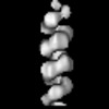 11847MC M: map data used to model this data C: citing same article ( |
|---|---|
| Similar structure data |
-Links
 PDBj
PDBj- Assembly
| Deposited unit | 
|
|---|---|
| 1 |
|
-Components
| #1: Protein | Mass: 26698.760 Da / Num. of mol.: 9 / Source method: isolated from a natural source / Source: (natural) Bunyamwera virus / Cell line: BHK-21 / References: UniProt: P16495 |
|---|
-Experimental details
-Experiment
| Experiment | Method: ELECTRON MICROSCOPY |
|---|---|
| EM experiment | Aggregation state: FILAMENT / 3D reconstruction method: single particle reconstruction |
- Sample preparation
| Component | Name: Native ribonucleoprotein extracted from Bunyamwera virus Type: COMPLEX / Entity ID: all / Source: NATURAL |
|---|---|
| Molecular weight | Experimental value: NO |
| Source (natural) | Organism: Bunyamwera virus |
| Buffer solution | pH: 7.4 |
| Specimen | Embedding applied: NO / Shadowing applied: NO / Staining applied: NO / Vitrification applied: YES |
| Vitrification | Instrument: FEI VITROBOT MARK II / Cryogen name: NITROGEN |
- Electron microscopy imaging
| Experimental equipment |  Model: Titan Krios / Image courtesy: FEI Company |
|---|---|
| Microscopy | Model: FEI TITAN KRIOS |
| Electron gun | Electron source:  FIELD EMISSION GUN / Accelerating voltage: 300 kV / Illumination mode: FLOOD BEAM FIELD EMISSION GUN / Accelerating voltage: 300 kV / Illumination mode: FLOOD BEAM |
| Electron lens | Mode: BRIGHT FIELD / Nominal magnification: 130000 X |
| Image recording | Average exposure time: 9 sec. / Electron dose: 51.3 e/Å2 / Detector mode: COUNTING / Film or detector model: GATAN K2 SUMMIT (4k x 4k) |
| EM imaging optics | Phase plate: VOLTA PHASE PLATE |
- Processing
| EM software |
| ||||||||||||
|---|---|---|---|---|---|---|---|---|---|---|---|---|---|
| CTF correction | Type: PHASE FLIPPING AND AMPLITUDE CORRECTION | ||||||||||||
| 3D reconstruction | Resolution: 13 Å / Resolution method: FSC 0.143 CUT-OFF / Num. of particles: 5077 / Symmetry type: POINT | ||||||||||||
| Atomic model building | Protocol: FLEXIBLE FIT | ||||||||||||
| Atomic model building | PDB-ID: 3ZLA Pdb chain-ID: B / Accession code: 3ZLA / Source name: PDB / Type: experimental model |

