 Movie
Movie Controller

[English] 日本語
 Yorodumi
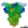
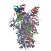
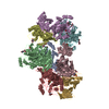
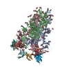
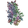
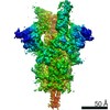
- PDB-7akd: Structure of the SARS-CoV-2 spike glycoprotein in complex with th... -
+ Open data

- Basic information
| Entry | Database: PDB / ID: 7akd | ||||||||||||||||||
|---|---|---|---|---|---|---|---|---|---|---|---|---|---|---|---|---|---|---|---|
| Title | Structure of the SARS-CoV-2 spike glycoprotein in complex with the 47D11 neutralizing antibody Fab fragment | ||||||||||||||||||
 Components Components |
| ||||||||||||||||||
 Keywords Keywords | VIRAL PROTEIN / SARS-coV-2 / spike / neutralizing antibody | ||||||||||||||||||
| Function / homology |  Function and homology information Function and homology informationsymbiont-mediated disruption of host tissue / Maturation of spike protein / Translation of Structural Proteins / Virion Assembly and Release / host cell surface / host extracellular region / symbiont-mediated-mediated suppression of host tetherin activity / Induction of Cell-Cell Fusion / structural constituent of virion / positive regulation of viral entry into host cell ...symbiont-mediated disruption of host tissue / Maturation of spike protein / Translation of Structural Proteins / Virion Assembly and Release / host cell surface / host extracellular region / symbiont-mediated-mediated suppression of host tetherin activity / Induction of Cell-Cell Fusion / structural constituent of virion / positive regulation of viral entry into host cell / membrane fusion / host cell endoplasmic reticulum-Golgi intermediate compartment membrane / Attachment and Entry / entry receptor-mediated virion attachment to host cell / receptor-mediated virion attachment to host cell / host cell surface receptor binding / symbiont-mediated suppression of host innate immune response / endocytosis involved in viral entry into host cell / receptor ligand activity / fusion of virus membrane with host plasma membrane / fusion of virus membrane with host endosome membrane / viral envelope / symbiont entry into host cell / virion attachment to host cell / host cell plasma membrane / SARS-CoV-2 activates/modulates innate and adaptive immune responses / virion membrane / membrane / identical protein binding / plasma membrane Similarity search - Function | ||||||||||||||||||
| Biological species |   Severe acute respiratory syndrome coronavirus 2 Severe acute respiratory syndrome coronavirus 2 Homo sapiens (human) Homo sapiens (human) | ||||||||||||||||||
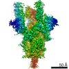
| Method | ELECTRON MICROSCOPY / single particle reconstruction / cryo EM / Resolution: 4 Å | ||||||||||||||||||
 Authors Authors | Fedry, J. / Hurdiss, D.L. / Wang, C. / Li, W. / Obal, G. / Drulyte, I. / Howes, S.C. / van Kuppeveld, F.J.M. / Foerster, F. / Bosch, B.J. | ||||||||||||||||||
| Funding support |  Netherlands, 5items Netherlands, 5items
| ||||||||||||||||||
 Citation Citation | Journal: Sci Adv / Year: 2021 Title: Structural insights into the cross-neutralization of SARS-CoV and SARS-CoV-2 by the human monoclonal antibody 47D11. Authors: Juliette Fedry / Daniel L Hurdiss / Chunyan Wang / Wentao Li / Gonzalo Obal / Ieva Drulyte / Wenjuan Du / Stuart C Howes / Frank J M van Kuppeveld / Friedrich Förster / Berend-Jan Bosch / Abstract: The emergence of SARS-CoV-2 antibody escape mutations highlights the urgent need for broadly neutralizing therapeutics. We previously identified a human monoclonal antibody, 47D11, capable of cross- ...The emergence of SARS-CoV-2 antibody escape mutations highlights the urgent need for broadly neutralizing therapeutics. We previously identified a human monoclonal antibody, 47D11, capable of cross-neutralizing SARS-CoV-2 and SARS-CoV and protecting against the associated respiratory disease in an animal model. Here, we report cryo-EM structures of both trimeric spike ectodomains in complex with the 47D11 Fab. 47D11 binds to the closed receptor-binding domain, distal to the ACE2 binding site. The CDRL3 stabilizes the N343 glycan in an upright conformation, exposing a mutationally constrained hydrophobic pocket, into which the CDRH3 loop inserts two aromatic residues. 47D11 stabilizes a partially open conformation of the SARS-CoV-2 spike, suggesting that it could be used effectively in combination with other antibodies targeting the exposed receptor-binding motif. Together, these results reveal a cross-protective epitope on the SARS-CoV-2 spike and provide a structural roadmap for the development of 47D11 as a prophylactic or postexposure therapy for COVID-19. | ||||||||||||||||||
| History |
|
- Structure visualization
| Movie |
Movie viewer |
|---|---|
| Structure viewer | Molecule: MolmilJmol/JSmol |
- Downloads & links
-Download
| PDBx/mmCIF format | 7akd.cif.gz | 622.4 KB | Display | PDBx/mmCIF format |
|---|---|---|---|---|
| PDB format | pdb7akd.ent.gz | 503.8 KB | Display | PDB format |
| PDBx/mmJSON format | 7akd.json.gz | Tree view | PDBx/mmJSON format | |
| Others |  Other downloads Other downloads |
-Validation report
| Arichive directory | https://data.pdbj.org/pub/pdb/validation_reports/ak/7akdftp://data.pdbj.org/pub/pdb/validation_reports/ak/7akd | HTTPS FTP |
|---|
-Related structure data
| Related structure data | 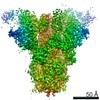 11812MC 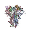 7akjC M: map data used to model this data C: citing same article ( |
|---|---|
| Similar structure data |
-Links
 PDBj
PDBj


- Assembly
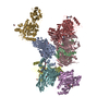
| Deposited unit | 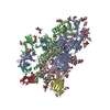
|
|---|---|
| 1 |
|
-Components
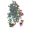
-Protein , 1 types, 3 molecules ABC
| #1: Protein | Mass: 141680.594 Da / Num. of mol.: 3 Source method: isolated from a genetically manipulated source Source: (gene. exp.) Severe acute respiratory syndrome coronavirus 2Gene: S, 2 / Plasmid: pCAGGS / Cell line (production host): 293 HEK F / Production host: Homo sapiens (human) / References: UniProt: P0DTC2 |
|---|
-Antibody , 2 types, 4 molecules DHEL
| #2: Antibody | Mass: 13205.812 Da / Num. of mol.: 2 Source method: isolated from a genetically manipulated source Source: (gene. exp.) Homo sapiens (human) / Plasmid: pFUSEss / Cell line (production host): 293 HEK F / Production host: Homo sapiens (human)#3: Antibody | Mass: 11350.607 Da / Num. of mol.: 2 Source method: isolated from a genetically manipulated source Source: (gene. exp.) Homo sapiens (human) / Plasmid: pFUSE2ss / Cell line (production host): 293 HEK F / Production host: Homo sapiens (human) |
|---|
-Sugars , 9 types, 50 molecules 


| #4: Polysaccharide | 2-acetamido-2-deoxy-beta-D-glucopyranose-(1-4)-2-acetamido-2-deoxy-beta-D-glucopyranose Source method: isolated from a genetically manipulated source #5: Polysaccharide | Source method: isolated from a genetically manipulated source #6: Polysaccharide | alpha-D-mannopyranose-(1-3)-[alpha-D-mannopyranose-(3-6)]beta-D-mannopyranose-(1-4)-2-acetamido-2- ...alpha-D-mannopyranose-(1-3)-[alpha-D-mannopyranose-(3-6)]beta-D-mannopyranose-(1-4)-2-acetamido-2-deoxy-beta-D-glucopyranose-(1-4)-2-acetamido-2-deoxy-beta-D-glucopyranose | Type: oligosaccharide / Mass: 910.823 Da / Num. of mol.: 1 Source method: isolated from a genetically manipulated source #7: Polysaccharide | beta-D-mannopyranose-(1-4)-2-acetamido-2-deoxy-beta-D-glucopyranose-(1-4)-[alpha-L-fucopyranose-(1- ...beta-D-mannopyranose-(1-4)-2-acetamido-2-deoxy-beta-D-glucopyranose-(1-4)-[alpha-L-fucopyranose-(1-6)]2-acetamido-2-deoxy-beta-D-glucopyranose | Source method: isolated from a genetically manipulated source #8: Polysaccharide | alpha-D-mannopyranose-(1-3)-alpha-D-mannopyranose-(1-6)-beta-D-mannopyranose-(1-4)-2-acetamido-2- ...alpha-D-mannopyranose-(1-3)-alpha-D-mannopyranose-(1-6)-beta-D-mannopyranose-(1-4)-2-acetamido-2-deoxy-beta-D-glucopyranose-(1-4)-[alpha-L-fucopyranose-(1-6)]2-acetamido-2-deoxy-beta-D-glucopyranose | #9: Polysaccharide | alpha-D-mannopyranose-(1-6)-alpha-D-mannopyranose | #10: Sugar | ChemComp-NAG /  Type: D-saccharide, beta linking / Mass: 221.208 Da / Num. of mol.: 28 / Source method: obtained synthetically / Formula: C8H15NO6 Type: D-saccharide, beta linking / Mass: 221.208 Da / Num. of mol.: 28 / Source method: obtained synthetically / Formula: C8H15NO6#11: Sugar |  Type: L-saccharide, alpha linking / Mass: 164.156 Da / Num. of mol.: 3 / Source method: obtained synthetically / Formula: C6H12O5 Type: L-saccharide, alpha linking / Mass: 164.156 Da / Num. of mol.: 3 / Source method: obtained synthetically / Formula: C6H12O5#12: Sugar |  Type: D-saccharide, alpha linking / Mass: 180.156 Da / Num. of mol.: 2 / Source method: obtained synthetically / Formula: C6H12O6 Type: D-saccharide, alpha linking / Mass: 180.156 Da / Num. of mol.: 2 / Source method: obtained synthetically / Formula: C6H12O6 |
|---|
-Details
| Has ligand of interest | N |
|---|---|
| Has protein modification | Y |
-Experimental details
-Experiment
| Experiment | Method: ELECTRON MICROSCOPY |
|---|---|
| EM experiment | Aggregation state: PARTICLE / 3D reconstruction method: single particle reconstruction |
- Sample preparation
| Component | Name: Complex of the SARS-CoV-2 spike glycoprotein with the 47D11 neutralizing antibody Fab fragment Type: COMPLEX / Entity ID: #1-#3 / Source: MULTIPLE SOURCES |
|---|---|
| Molecular weight | Value: 0.55 MDa / Experimental value: YES |
| Source (natural) | Organism: Homo sapiens (human) |
| Source (recombinant) | Organism: Homo sapiens (human) / Cell: HEK293F / Plasmid: pCAGGS |
| Buffer solution | pH: 8 |
| Specimen | Conc.: 1.6 mg/ml / Embedding applied: NO / Shadowing applied: NO / Staining applied: NO / Vitrification applied: YES |
| Specimen support | Grid material: COPPER / Grid type: Quantifoil R1.2/1.3 |
| Vitrification | Instrument: FEI VITROBOT MARK IV / Cryogen name: ETHANE / Humidity: 100 % / Chamber temperature: 277 K |
- Electron microscopy imaging
| Experimental equipment |  Model: Talos Arctica / Image courtesy: FEI Company |
|---|---|
| Microscopy | Model: FEI TALOS ARCTICA |
| Electron gun | Electron source:  FIELD EMISSION GUN / Accelerating voltage: 200 kV / Illumination mode: FLOOD BEAM FIELD EMISSION GUN / Accelerating voltage: 200 kV / Illumination mode: FLOOD BEAM |
| Electron lens | Mode: BRIGHT FIELD / Nominal magnification: 130000 X / Nominal defocus max: 2500 nm / Nominal defocus min: 500 nm / Cs: 2.7 mm / C2 aperture diameter: 30 µm |
| Specimen holder | Cryogen: NITROGEN / Specimen holder model: OTHER |
| Image recording | Electron dose: 40 e/Å2 / Detector mode: COUNTING / Film or detector model: GATAN K2 SUMMIT (4k x 4k) / Num. of real images: 4231 |
| EM imaging optics | Energyfilter name: GIF Quantum LS / Energyfilter slit width: 20 eV |
- Processing
| Software | Name: UCSF ChimeraX / Version: 0.93/v8 / Classification: model building / URL: https://www.rbvi.ucsf.edu/chimerax/ / Os: macOS / Type: package | ||||||||||||||||||||||||
|---|---|---|---|---|---|---|---|---|---|---|---|---|---|---|---|---|---|---|---|---|---|---|---|---|---|
| EM software |
| ||||||||||||||||||||||||
| CTF correction | Type: PHASE FLIPPING AND AMPLITUDE CORRECTION | ||||||||||||||||||||||||
| Particle selection | Num. of particles selected: 1500537 | ||||||||||||||||||||||||
| 3D reconstruction | Resolution: 4 Å / Resolution method: FSC 0.143 CUT-OFF / Num. of particles: 945232 / Symmetry type: POINT |
