 Movie
Movie Controller

[English] 日本語
 Yorodumi
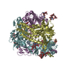
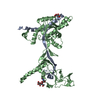
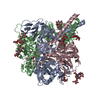
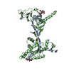
- PDB-6xye: Cryo-EM structure of the prefusion state of canine distemper viru... -
+ Open data

- Basic information
| Entry | Database: PDB / ID: 6xye | ||||||
|---|---|---|---|---|---|---|---|
| Title | Cryo-EM structure of the prefusion state of canine distemper virus fusion protein ectodomain | ||||||
 Components Components |
| ||||||
 Keywords Keywords | VIRAL PROTEIN / fusion protein / prefusion state | ||||||
| Function / homology |  Function and homology information Function and homology informationfusion of virus membrane with host plasma membrane / viral envelope / symbiont entry into host cell / host cell plasma membrane / virion membrane / plasma membrane Similarity search - Function | ||||||
| Biological species |  Canine morbillivirus Canine morbillivirus | ||||||
| Method | ELECTRON MICROSCOPY / single particle reconstruction / cryo EM / Resolution: 4.3 Å | ||||||
 Authors Authors | Kalbermatter, D. / Fotiadis, D. | ||||||
| Funding support |  Switzerland, 1items Switzerland, 1items
| ||||||
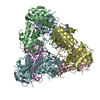
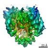
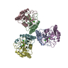
 Citation Citation | Journal: J Struct Biol X / Year: 2020 Title: Cryo-EM structure of the prefusion state of canine distemper virus fusion protein ectodomain. Authors: David Kalbermatter / Neeta Shrestha / Flavio M Gall / Marianne Wyss / Rainer Riedl / Philippe Plattet / Dimitrios Fotiadis / Abstract: Measles virus (MeV) and canine distemper virus (CDV), two members of the genus, are still causing important global diseases of humans and animals, respectively. To enter target cells, ...Measles virus (MeV) and canine distemper virus (CDV), two members of the genus, are still causing important global diseases of humans and animals, respectively. To enter target cells, morbilliviruses rely on an envelope-anchored machinery, which is composed of two interacting glycoproteins: a tetrameric receptor binding (H) protein and a trimeric fusion (F) protein. To execute membrane fusion, the F protein initially adopts a metastable, prefusion state that refolds into a highly stable postfusion conformation as the result of a finely coordinated activation process mediated by the H protein. Here, we employed cryo-electron microscopy (cryo-EM) and single particle reconstruction to elucidate the structure of the prefusion state of the CDV F protein ectodomain (solF) at 4.3 Å resolution. Stabilization of the prefusion solF trimer was achieved by fusing the GCNt trimerization sequence at the C-terminal protein region, and expressing and purifying the recombinant protein in the presence of a morbilliviral fusion inhibitor class compound. The three-dimensional cryo-EM map of prefusion CDV solF in complex with the inhibitor clearly shows density for the ligand at the protein binding site suggesting common mechanisms of membrane fusion activation and inhibition employed by different morbillivirus members. | ||||||
| History |
|
- Structure visualization
| Movie |
Movie viewer |
|---|---|
| Structure viewer | Molecule: MolmilJmol/JSmol |
- Downloads & links
-Download
| PDBx/mmCIF format | 6xye.cif.gz | 226.5 KB | Display | PDBx/mmCIF format |
|---|---|---|---|---|
| PDB format | pdb6xye.ent.gz | 180.2 KB | Display | PDB format |
| PDBx/mmJSON format | 6xye.json.gz | Tree view | PDBx/mmJSON format | |
| Others |  Other downloads Other downloads |
-Validation report
| Arichive directory | https://data.pdbj.org/pub/pdb/validation_reports/xy/6xyeftp://data.pdbj.org/pub/pdb/validation_reports/xy/6xye | HTTPS FTP |
|---|
-Related structure data
| Related structure data | 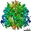 10649MC M: map data used to model this data C: citing same article ( |
|---|---|
| Similar structure data | |
| EM raw data | EMPIAR-10366 (Title: Cryo-EM structure of the prefusion state of canine distemper virus fusion protein ectodomain Data size: 474.1 Data #1: Unaligned multi-frame micrographs of the prefusion state of canine distemper virus fusion protein ectodomain. [micrographs - multiframe]) |
-Links
 PDBj
PDBj

- Assembly
| Deposited unit | 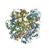
|
|---|---|
| 1 |
|
-Components
| #1: Protein | Mass: 10128.781 Da / Num. of mol.: 3 Source method: isolated from a genetically manipulated source Source: (gene. exp.) Canine morbillivirus / Cell line (production host): HEK293 / Production host:  Homo sapiens (human) / References: UniProt: Q9YKL7, UniProt: A0A0R5ZPI3*PLUS Homo sapiens (human) / References: UniProt: Q9YKL7, UniProt: A0A0R5ZPI3*PLUS#2: Protein | Mass: 47511.637 Da / Num. of mol.: 3 Source method: isolated from a genetically manipulated source Source: (gene. exp.) Canine morbillivirus / Cell line (production host): HEK293 / Production host: Homo sapiens (human) / References: UniProt: Q9YKL7Has protein modification | Y | |
|---|
-Experimental details
-Experiment
| Experiment | Method: ELECTRON MICROSCOPY |
|---|---|
| EM experiment | Aggregation state: PARTICLE / 3D reconstruction method: single particle reconstruction |
- Sample preparation
| Component | Name: Canine distemper virus fusion protein / Type: COMPLEX / Entity ID: all / Source: RECOMBINANT | |||||||||||||||||||||||||
|---|---|---|---|---|---|---|---|---|---|---|---|---|---|---|---|---|---|---|---|---|---|---|---|---|---|---|
| Source (natural) | Organism: Canine morbillivirus | |||||||||||||||||||||||||
| Source (recombinant) | Organism: Homo sapiens (human) / Cell: HEK293 | |||||||||||||||||||||||||
| Buffer solution | pH: 7.5 | |||||||||||||||||||||||||
| Buffer component |
| |||||||||||||||||||||||||
| Specimen | Conc.: 0.23 mg/ml / Embedding applied: NO / Shadowing applied: NO / Staining applied: NO / Vitrification applied: YES | |||||||||||||||||||||||||
| Specimen support | Grid material: COPPER / Grid mesh size: 200 divisions/in. / Grid type: Quantifoil R2/1 | |||||||||||||||||||||||||
| Vitrification | Instrument: FEI VITROBOT MARK IV / Cryogen name: ETHANE / Humidity: 100 % / Chamber temperature: 277.15 K |
- Electron microscopy imaging
| Experimental equipment |  Model: Tecnai Polara / Image courtesy: FEI Company |
|---|---|
| Microscopy | Model: FEI POLARA 300 |
| Electron gun | Electron source:  FIELD EMISSION GUN / Accelerating voltage: 300 kV / Illumination mode: FLOOD BEAM FIELD EMISSION GUN / Accelerating voltage: 300 kV / Illumination mode: FLOOD BEAM |
| Electron lens | Mode: BRIGHT FIELD |
| Specimen holder | Cryogen: NITROGEN |
| Image recording | Electron dose: 73 e/Å2 / Detector mode: COUNTING / Film or detector model: GATAN K2 SUMMIT (4k x 4k) / Num. of grids imaged: 1 / Num. of real images: 1604 |
| Image scans | Movie frames/image: 48 |
- Processing
| EM software |
| |||||||||||||||||||||||||||||||||||||||||||||||||||||||
|---|---|---|---|---|---|---|---|---|---|---|---|---|---|---|---|---|---|---|---|---|---|---|---|---|---|---|---|---|---|---|---|---|---|---|---|---|---|---|---|---|---|---|---|---|---|---|---|---|---|---|---|---|---|---|---|---|
| CTF correction | Type: PHASE FLIPPING AND AMPLITUDE CORRECTION | |||||||||||||||||||||||||||||||||||||||||||||||||||||||
| Particle selection | Num. of particles selected: 491315 | |||||||||||||||||||||||||||||||||||||||||||||||||||||||
| Symmetry | Point symmetry: C3 (3 fold cyclic) | |||||||||||||||||||||||||||||||||||||||||||||||||||||||
| 3D reconstruction | Resolution: 4.3 Å / Resolution method: FSC 0.143 CUT-OFF / Num. of particles: 115248 / Num. of class averages: 3 / Symmetry type: POINT | |||||||||||||||||||||||||||||||||||||||||||||||||||||||
| Atomic model building | Protocol: RIGID BODY FIT / Space: REAL | |||||||||||||||||||||||||||||||||||||||||||||||||||||||
| Atomic model building | 3D fitting-ID: 1 / Accession code: 5YZC / Initial refinement model-ID: 1 / PDB-ID: 5YZC / Source name: PDB / Type: experimental model
|

