 Movie
Movie Controller


+ Open data

- Basic information
| Entry | Database: PDB / ID: 6w24 | ||||||
|---|---|---|---|---|---|---|---|
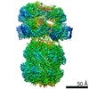
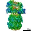
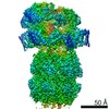
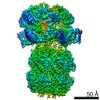
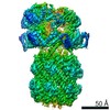
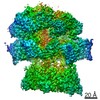
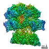
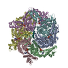
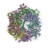
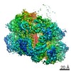
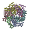
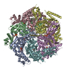
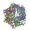
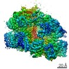
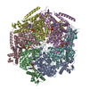
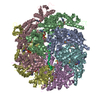
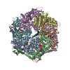
| Title | ClpA Engaged2 State bound to RepA-GFP (Focused Classification) | ||||||
 Components Components |
| ||||||
 Keywords Keywords | CHAPERONE / AAA+ / Protease / Hsp100 / ATPase | ||||||
| Function / homology |  Function and homology information Function and homology informationendopeptidase Clp complex / ATP-dependent peptidase activity / protein quality control for misfolded or incompletely synthesized proteins / protein unfolding / cellular response to heat / response to oxidative stress / ATP hydrolysis activity / proteolysis / ATP binding / cytosol / cytoplasm Similarity search - Function | ||||||
| Biological species |  synthetic construct (others) | ||||||
| Method | ELECTRON MICROSCOPY / single particle reconstruction / cryo EM / Resolution: 3.4 Å | ||||||
 Authors Authors | Lopez, K.L. / Rizo, A.N. / Tse, E. / Lin, J. / Scull, N.W. / Thwin, A.C. / Lucius, A.L. / Shorter, J. / Southworth, D.R. | ||||||
| Funding support |  United States, 1items United States, 1items
| ||||||
 Citation Citation | Journal: Nat Struct Mol Biol / Year: 2020 Title: Conformational plasticity of the ClpAP AAA+ protease couples protein unfolding and proteolysis. Authors: Kyle E Lopez / Alexandrea N Rizo / Eric Tse / JiaBei Lin / Nathaniel W Scull / Aye C Thwin / Aaron L Lucius / James Shorter / Daniel R Southworth / Abstract: The ClpAP complex is a conserved bacterial protease that unfolds and degrades proteins targeted for destruction. The ClpA double-ring hexamer powers substrate unfolding and translocation into the ...The ClpAP complex is a conserved bacterial protease that unfolds and degrades proteins targeted for destruction. The ClpA double-ring hexamer powers substrate unfolding and translocation into the ClpP proteolytic chamber. Here, we determined high-resolution structures of wild-type Escherichia coli ClpAP undergoing active substrate unfolding and proteolysis. A spiral of pore loop-substrate contacts spans both ClpA AAA+ domains. Protomers at the spiral seam undergo nucleotide-specific rearrangements, supporting substrate translocation. IGL loops extend flexibly to bind the planar, heptameric ClpP surface with the empty, symmetry-mismatched IGL pocket maintained at the seam. Three different structures identify a binding-pocket switch by the IGL loop of the lowest positioned protomer, involving release and re-engagement with the clockwise pocket. This switch is coupled to a ClpA rotation and a network of conformational changes across the seam, suggesting that ClpA can rotate around the ClpP apical surface during processive steps of translocation and proteolysis. | ||||||
| History |
|
- Structure visualization
| Movie |
Movie viewer |
|---|---|
| Structure viewer | Molecule: MolmilJmol/JSmol |
- Downloads & links
-Download
| PDBx/mmCIF format | 6w24.cif.gz | 596.3 KB | Display | PDBx/mmCIF format |
|---|---|---|---|---|
| PDB format | pdb6w24.ent.gz | 493.6 KB | Display | PDB format |
| PDBx/mmJSON format | 6w24.json.gz | Tree view | PDBx/mmJSON format | |
| Others |  Other downloads Other downloads |
-Validation report
| Summary document | 6w24_validation.pdf.gz | 1.7 MB | Display | wwPDB validaton report |
|---|---|---|---|---|
| Full document | 6w24_full_validation.pdf.gz | 1.8 MB | Display | |
| Data in XML | 6w24_validation.xml.gz | 105 KB | Display | |
| Data in CIF | 6w24_validation.cif.gz | 150.6 KB | Display | |
| Arichive directory | https://data.pdbj.org/pub/pdb/validation_reports/w2/6w24ftp://data.pdbj.org/pub/pdb/validation_reports/w2/6w24 | HTTPS FTP |
-Related structure data
| Related structure data |  21524MC 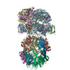 6uqeC 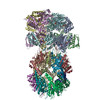 6uqoC 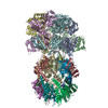 6w1zC 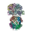 6w20C 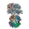 6w21C 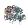 6w22C 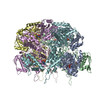 6w23C M: map data used to model this data C: citing same article ( |
|---|---|
| Similar structure data |
-Links
 PDBj
PDBj


- Assembly
| Deposited unit | 
|
|---|---|
| 1 |
|
-Components
| #1: Protein | Mass: 84321.844 Da / Num. of mol.: 6 Source method: isolated from a genetically manipulated source Source: (gene. exp.) Strain: K12 / Gene: clpA, lopD, b0882, JW0866 / Production host: #2: Protein/peptide | | Mass: 2060.531 Da / Num. of mol.: 1 Source method: isolated from a genetically manipulated source Source: (gene. exp.) synthetic construct (others) / Production host: #3: Chemical | ChemComp-ADP /   Mass: 427.201 Da / Num. of mol.: 5 / Source method: obtained synthetically / Formula: C10H15N5O10P2 / Comment: ADP, energy-carrying molecule*YM Mass: 427.201 Da / Num. of mol.: 5 / Source method: obtained synthetically / Formula: C10H15N5O10P2 / Comment: ADP, energy-carrying molecule*YM#4: Chemical | ChemComp-ATP /   Mass: 507.181 Da / Num. of mol.: 7 / Source method: obtained synthetically / Formula: C10H16N5O13P3 / Comment: ATP, energy-carrying molecule*YM Mass: 507.181 Da / Num. of mol.: 7 / Source method: obtained synthetically / Formula: C10H16N5O13P3 / Comment: ATP, energy-carrying molecule*YMHas ligand of interest | N | |
|---|
-Experimental details
-Experiment
| Experiment | Method: ELECTRON MICROSCOPY |
|---|---|
| EM experiment | Aggregation state: PARTICLE / 3D reconstruction method: single particle reconstruction |
- Sample preparation
| Component |
| ||||||||||||||||||||||||
|---|---|---|---|---|---|---|---|---|---|---|---|---|---|---|---|---|---|---|---|---|---|---|---|---|---|
| Molecular weight | Value: 0.84 MDa / Experimental value: NO | ||||||||||||||||||||||||
| Source (natural) |
| ||||||||||||||||||||||||
| Source (recombinant) |
| ||||||||||||||||||||||||
| Buffer solution | pH: 7.5 | ||||||||||||||||||||||||
| Specimen | Embedding applied: NO / Shadowing applied: NO / Staining applied: NO / Vitrification applied: YES | ||||||||||||||||||||||||
| Vitrification | Cryogen name: ETHANE |
- Electron microscopy imaging
| Experimental equipment |  Model: Titan Krios / Image courtesy: FEI Company |
|---|---|
| Microscopy | Model: FEI TITAN KRIOS |
| Electron gun | Electron source:  FIELD EMISSION GUN / Accelerating voltage: 300 kV / Illumination mode: FLOOD BEAM FIELD EMISSION GUN / Accelerating voltage: 300 kV / Illumination mode: FLOOD BEAM |
| Electron lens | Mode: BRIGHT FIELD / Nominal magnification: 105000 X / Calibrated magnification: 58616 X / Nominal defocus max: 2000 nm / Nominal defocus min: 1000 nm / Cs: 2.7 mm / C2 aperture diameter: 70 µm |
| Specimen holder | Cryogen: NITROGEN / Specimen holder model: FEI TITAN KRIOS AUTOGRID HOLDER |
| Image recording | Average exposure time: 6 sec. / Electron dose: 69 e/Å2 / Film or detector model: GATAN K3 BIOQUANTUM (6k x 4k) |
| Image scans | Width: 11520 / Height: 8184 |
- Processing
| Software | Name: PHENIX / Version: 1.17_3644: / Classification: refinement | ||||||||||||||||||||||||
|---|---|---|---|---|---|---|---|---|---|---|---|---|---|---|---|---|---|---|---|---|---|---|---|---|---|
| EM software | Name: cryoSPARC / Version: 2 / Category: 3D reconstruction | ||||||||||||||||||||||||
| CTF correction | Type: PHASE FLIPPING AND AMPLITUDE CORRECTION | ||||||||||||||||||||||||
| Symmetry | Point symmetry: C1 (asymmetric) | ||||||||||||||||||||||||
| 3D reconstruction | Resolution: 3.4 Å / Resolution method: FSC 0.143 CUT-OFF / Num. of particles: 39177 / Symmetry type: POINT | ||||||||||||||||||||||||
| Refine LS restraints |
|
