 Movie
Movie Controller

[English] 日本語
 Yorodumi
- EMDB-21523: ClpA Disengaged State bound to RepA-GFP (Focused Classification) -
+ Open data

- Basic information
| Entry | Database: EMDB / ID: EMD-21523 | |||||||||
|---|---|---|---|---|---|---|---|---|---|---|
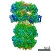
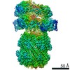
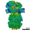
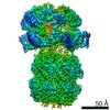
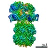
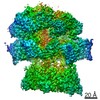
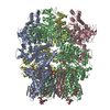
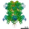
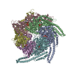
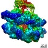
| Title | ClpA Disengaged State bound to RepA-GFP (Focused Classification) | |||||||||
 Map data Map data | ClpA Disengaged State bound to RepA-GFP (Focused Classification) | |||||||||
 Sample Sample |
| |||||||||
 Keywords Keywords | AAA+ / Chaperone / Protease / Hsp100 / ATPase | |||||||||
| Function / homology |  Function and homology information Function and homology informationendopeptidase Clp complex / ATP-dependent peptidase activity / protein quality control for misfolded or incompletely synthesized proteins / protein unfolding / cellular response to heat / response to oxidative stress / ATP hydrolysis activity / proteolysis / ATP binding / cytoplasm / cytosol Similarity search - Function | |||||||||
| Biological species |  | |||||||||
| Method | single particle reconstruction / cryo EM / Resolution: 3.1 Å | |||||||||
 Authors Authors | Lopez KL / Rizo AN | |||||||||
| Funding support |  United States, 1 items United States, 1 items
| |||||||||
 Citation Citation | Journal: Nat Struct Mol Biol / Year: 2020 Title: Conformational plasticity of the ClpAP AAA+ protease couples protein unfolding and proteolysis. Authors: Kyle E Lopez / Alexandrea N Rizo / Eric Tse / JiaBei Lin / Nathaniel W Scull / Aye C Thwin / Aaron L Lucius / James Shorter / Daniel R Southworth / Abstract: The ClpAP complex is a conserved bacterial protease that unfolds and degrades proteins targeted for destruction. The ClpA double-ring hexamer powers substrate unfolding and translocation into the ...The ClpAP complex is a conserved bacterial protease that unfolds and degrades proteins targeted for destruction. The ClpA double-ring hexamer powers substrate unfolding and translocation into the ClpP proteolytic chamber. Here, we determined high-resolution structures of wild-type Escherichia coli ClpAP undergoing active substrate unfolding and proteolysis. A spiral of pore loop-substrate contacts spans both ClpA AAA+ domains. Protomers at the spiral seam undergo nucleotide-specific rearrangements, supporting substrate translocation. IGL loops extend flexibly to bind the planar, heptameric ClpP surface with the empty, symmetry-mismatched IGL pocket maintained at the seam. Three different structures identify a binding-pocket switch by the IGL loop of the lowest positioned protomer, involving release and re-engagement with the clockwise pocket. This switch is coupled to a ClpA rotation and a network of conformational changes across the seam, suggesting that ClpA can rotate around the ClpP apical surface during processive steps of translocation and proteolysis. | |||||||||
| History |
|
- Structure visualization
| Movie |
Movie viewer |
|---|---|
| Structure viewer | EM map: SurfViewMolmilJmol/JSmol |
| Supplemental images |

- Downloads & links
-EMDB archive
| Map data | emd_21523.map.gz | 778.3 MB | EMDB map data format | |
|---|---|---|---|---|
| Header (meta data) | emd-21523-v30.xmlemd-21523.xml | 13.9 KB 13.9 KB | Display Display | EMDB header |
| Images | 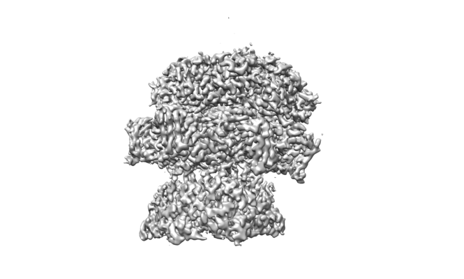 emd_21523.png emd_21523.png | 57.3 KB | ||
| Filedesc metadata | emd-21523.cif.gz | 5.8 KB | ||
| Archive directory |  http://ftp.pdbj.org/pub/emdb/structures/EMD-21523ftp://ftp.pdbj.org/pub/emdb/structures/EMD-21523 http://ftp.pdbj.org/pub/emdb/structures/EMD-21523ftp://ftp.pdbj.org/pub/emdb/structures/EMD-21523 | HTTPS FTP |
-Related structure data
| Related structure data |  6w23MC  6uqeC  6uqoC 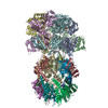 6w1zC 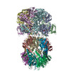 6w20C 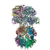 6w21C 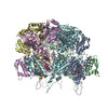 6w22C 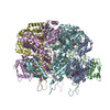 6w24C M: atomic model generated by this map C: citing same article ( |
|---|---|
| Similar structure data |


-Links
| EMDB pages | EMDB (EBI/PDBe) / EMDataResource |
|---|---|
| Related items in Molecule of the Month |


-Map
| File | Download / File: emd_21523.map.gz / Format: CCP4 / Size: 824 MB / Type: IMAGE STORED AS FLOATING POINT NUMBER (4 BYTES) | ||||||||||||||||||||||||||||||||||||||||||||||||||||||||||||
|---|---|---|---|---|---|---|---|---|---|---|---|---|---|---|---|---|---|---|---|---|---|---|---|---|---|---|---|---|---|---|---|---|---|---|---|---|---|---|---|---|---|---|---|---|---|---|---|---|---|---|---|---|---|---|---|---|---|---|---|---|---|
| Annotation | ClpA Disengaged State bound to RepA-GFP (Focused Classification) | ||||||||||||||||||||||||||||||||||||||||||||||||||||||||||||
| Projections & slices | Image control
Images are generated by Spider. | ||||||||||||||||||||||||||||||||||||||||||||||||||||||||||||
| Voxel size | X=Y=Z: 0.834 Å | ||||||||||||||||||||||||||||||||||||||||||||||||||||||||||||
| Density |
| ||||||||||||||||||||||||||||||||||||||||||||||||||||||||||||
| Symmetry | Space group: 1 | ||||||||||||||||||||||||||||||||||||||||||||||||||||||||||||
| Details | EMDB XML:
CCP4 map header:
| ||||||||||||||||||||||||||||||||||||||||||||||||||||||||||||
 Z (Sec.)
 Y (Row.)
 X (Col.)


-Supplemental data
- Sample components
-Entire : ClpAP
| Entire | Name: ClpAP |
|---|---|
| Components |
|
-Supramolecule #1: ClpAP
| Supramolecule | Name: ClpAP / type: complex / ID: 1 / Parent: 0 / Macromolecule list: #1-#2 |
|---|---|
| Molecular weight | Theoretical: 840 KDa |
-Supramolecule #2: ATP-dependent Clp protease ATP-binding subunit ClpA
| Supramolecule | Name: ATP-dependent Clp protease ATP-binding subunit ClpA / type: complex / ID: 2 / Parent: 1 / Macromolecule list: #1 |
|---|---|
| Source (natural) | Organism: |
-Supramolecule #3: RepA, green fluorescent protein fusion
| Supramolecule | Name: RepA, green fluorescent protein fusion / type: complex / ID: 3 / Parent: 1 / Macromolecule list: #2 |
|---|---|
| Source (natural) | Organism: synthetic construct (others) |
-Macromolecule #1: ATP-dependent Clp protease ATP-binding subunit ClpA
| Macromolecule | Name: ATP-dependent Clp protease ATP-binding subunit ClpA / type: protein_or_peptide / ID: 1 / Number of copies: 6 / Enantiomer: LEVO |
|---|---|
| Source (natural) | Organism: |
| Molecular weight | Theoretical: 84.321844 KDa |
| Recombinant expression | Organism: |
| Sequence | String: MLNQELELSL NMAFARAREH RHEFMTVEHL LLALLSNPSA REALEACSVD LVALRQELEA FIEQTTPVLP ASEEERDTQP TLSFQRVLQ RAVFHVQSSG RNEVTGANVL VAIFSEQESQ AAYLLRKHEV SRLDVVNFIS HGTRKDEPTQ SSDPGSQPNS E EQAGGEER ...String: MLNQELELSL NMAFARAREH RHEFMTVEHL LLALLSNPSA REALEACSVD LVALRQELEA FIEQTTPVLP ASEEERDTQP TLSFQRVLQ RAVFHVQSSG RNEVTGANVL VAIFSEQESQ AAYLLRKHEV SRLDVVNFIS HGTRKDEPTQ SSDPGSQPNS E EQAGGEER MENFTTNLNQ LARVGGIDPL IGREKELERA IQVLCRRRKN NPLLVGESGV GKTAIAEGLA WRIVQGDVPE VM ADCTIYS LDIGSLLAGT KYRGDFEKRF KALLKQLEQD TNSILFIDEI HTIIGAGAAS GGQVDAANLI KPLLSSGKIR VIG STTYQE FSNIFEKDRA LARRFQKIDI TEPSIEETVQ IINGLKPKYE AHHDVRYTAK AVRAAVELAV KYINDRHLPD KAID VIDEA GARARLMPVS KRKKTVNVAD IESVVARIAR IPEKSVSQSD RDTLKNLGDR LKMLVFGQDK AIEALTEAIK MARAG LGHE HKPVGSFLFA GPTGVGKTEV TVQLSKALGI ELLRFDMSEY MERHTVSRLI GAPPGYVGFD QGGLLTDAVI KHPHAV LLL DEIEKAHPDV FNILLQVMDN GTLTDNNGRK ADFRNVVLVM TTNAGVRETE RKSIGLIHQD NSTDAMEEIK KIFTPEF RN RLDNIIWFDH LSTDVIHQVV DKFIVELQVQ LDQKGVSLEV SQEARNWLAE KGYDRAMGAR PMARVIQDNL KKPLANEL L FGSLVDGGQV TVALDKEKNE LTYGFQSAQK HKAEAAH UniProtKB: ATP-dependent Clp protease ATP-binding subunit ClpA |
-Macromolecule #2: RepA, green fluorescent protein fusion
| Macromolecule | Name: RepA, green fluorescent protein fusion / type: protein_or_peptide / ID: 2 / Number of copies: 1 / Enantiomer: LEVO |
|---|---|
| Source (natural) | Organism: synthetic construct (others) |
| Molecular weight | Theoretical: 2.060531 KDa |
| Recombinant expression | Organism: |
| Sequence | String: (UNK)(UNK)(UNK)(UNK)(UNK)(UNK)(UNK)(UNK)(UNK)(UNK) (UNK)(UNK)(UNK)(UNK)(UNK)(UNK) (UNK)(UNK)(UNK) (UNK)(UNK)(UNK)(UNK)(UNK) |
-Macromolecule #3: ADENOSINE-5'-DIPHOSPHATE
| Macromolecule | Name: ADENOSINE-5'-DIPHOSPHATE / type: ligand / ID: 3 / Number of copies: 3 / Formula: ADP |
|---|---|
| Molecular weight | Theoretical: 427.201 Da |
| Chemical component information |  ChemComp-ADP: |
-Macromolecule #4: ADENOSINE-5'-TRIPHOSPHATE
| Macromolecule | Name: ADENOSINE-5'-TRIPHOSPHATE / type: ligand / ID: 4 / Number of copies: 8 / Formula: ATP |
|---|---|
| Molecular weight | Theoretical: 507.181 Da |
| Chemical component information |  ChemComp-ATP: |
-Experimental details
-Structure determination
| Method | cryo EM |
|---|---|
 Processing Processing | single particle reconstruction |
| Aggregation state | particle |
-Sample preparation
| Buffer | pH: 7.5 |
|---|---|
| Vitrification | Cryogen name: ETHANE |
- Electron microscopy
| Microscope | FEI TITAN KRIOS |
|---|---|
| Image recording | Film or detector model: GATAN K3 BIOQUANTUM (6k x 4k) / Digitization - Dimensions - Width: 11520 pixel / Digitization - Dimensions - Height: 8184 pixel / Average exposure time: 6.0 sec. / Average electron dose: 69.0 e/Å2 |
| Electron beam | Acceleration voltage: 300 kV / Electron source:  FIELD EMISSION GUN FIELD EMISSION GUN |
| Electron optics | C2 aperture diameter: 70.0 µm / Calibrated magnification: 58616 / Illumination mode: FLOOD BEAM / Imaging mode: BRIGHT FIELD / Cs: 2.7 mm / Nominal defocus max: 2.0 µm / Nominal defocus min: 1.0 µm / Nominal magnification: 105000 |
| Sample stage | Specimen holder model: FEI TITAN KRIOS AUTOGRID HOLDER / Cooling holder cryogen: NITROGEN |
| Experimental equipment |  Model: Titan Krios / Image courtesy: FEI Company |
-Image processing
| Startup model | Type of model: INSILICO MODEL |
|---|---|
| Final reconstruction | Applied symmetry - Point group: C1 (asymmetric) / Resolution.type: BY AUTHOR / Resolution: 3.1 Å / Resolution method: FSC 0.143 CUT-OFF / Software - Name: cryoSPARC (ver. 2) / Number images used: 57848 |
| Initial angle assignment | Type: PROJECTION MATCHING |
| Final angle assignment | Type: PROJECTION MATCHING |
