 Movie
Movie Controller


+ Open data

- Basic information
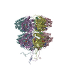
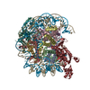
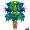
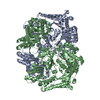
| Entry | Database: PDB / ID: 6v1s | |||||||||||||||||||||||||||||||||
|---|---|---|---|---|---|---|---|---|---|---|---|---|---|---|---|---|---|---|---|---|---|---|---|---|---|---|---|---|---|---|---|---|---|---|
| Title | Structure of the Clostridioides difficile transferase toxin | |||||||||||||||||||||||||||||||||
 Components Components |
| |||||||||||||||||||||||||||||||||
 Keywords Keywords | TRANSLOCASE / Clostridium / Clostridioides / Binary / CDT / Iota / Toxin | |||||||||||||||||||||||||||||||||
| Function / homology |  Function and homology information Function and homology informationglycosyltransferase activity / Transferases; Glycosyltransferases; Pentosyltransferases / protein homooligomerization / transferase activity / nucleotide binding / extracellular region / metal ion binding / identical protein binding Similarity search - Function | |||||||||||||||||||||||||||||||||
| Biological species |  Clostridioides difficile (bacteria) Clostridioides difficile (bacteria) | |||||||||||||||||||||||||||||||||
| Method | ELECTRON MICROSCOPY / single particle reconstruction / cryo EM / Resolution: 3.8 Å | |||||||||||||||||||||||||||||||||
 Authors Authors | Sheedlo, M.J. / Anderson, D.M. / Thomas, A.K. / Lacy, D.B. | |||||||||||||||||||||||||||||||||
| Funding support |  United States, 2items United States, 2items
| |||||||||||||||||||||||||||||||||
 Citation Citation | Journal: Proc Natl Acad Sci U S A / Year: 2020 Title: Structural elucidation of the transferase toxin reveals a single-site binding mode for the enzyme. Authors: Michael J Sheedlo / David M Anderson / Audrey K Thomas / D Borden Lacy / Abstract: is a Gram-positive, pathogenic bacterium and a prominent cause of hospital-acquired diarrhea in the United States. The symptoms of infection are caused by the activity of three large toxins known ... is a Gram-positive, pathogenic bacterium and a prominent cause of hospital-acquired diarrhea in the United States. The symptoms of infection are caused by the activity of three large toxins known as toxin A (TcdA), toxin B (TcdB), and the transferase toxin (CDT). Reported here is a 3.8-Å cryo-electron microscopy (cryo-EM) structure of CDT, a bipartite toxin comprised of the proteins CDTa and CDTb. We observe a single molecule of CDTa bound to a CDTb heptamer. The formation of the CDT complex relies on the interaction of an N-terminal adaptor and pseudoenzyme domain of CDTa with six subunits of the CDTb heptamer. CDTb is observed in a preinsertion state, a conformation observed in the transition of prepore to β-barrel pore, although we also observe a single bound CDTa in the prepore and β-barrel conformations of CDTb. The binding interaction appears to prime CDTa for translocation as the adaptor subdomain enters the lumen of the preinsertion state channel. These structural observations advance the understanding of how a single protein, CDTb, can mediate the delivery of a large enzyme, CDTa, into the cytosol of mammalian cells. | |||||||||||||||||||||||||||||||||
| History |
|
- Structure visualization
| Movie |
Movie viewer |
|---|---|
| Structure viewer | Molecule: MolmilJmol/JSmol |
- Downloads & links
-Download
| PDBx/mmCIF format | 6v1s.cif.gz | 476.2 KB | Display | PDBx/mmCIF format |
|---|---|---|---|---|
| PDB format | pdb6v1s.ent.gz | 341.7 KB | Display | PDB format |
| PDBx/mmJSON format | 6v1s.json.gz | Tree view | PDBx/mmJSON format | |
| Others |  Other downloads Other downloads |
-Validation report
| Arichive directory | https://data.pdbj.org/pub/pdb/validation_reports/v1/6v1sftp://data.pdbj.org/pub/pdb/validation_reports/v1/6v1s | HTTPS FTP |
|---|
-Related structure data
| Related structure data |  21016MC M: map data used to model this data C: citing same article ( |
|---|---|
| Similar structure data |


-Links
 PDBj
PDBj
- Assembly
| Deposited unit | 
|
|---|---|
| 1 |
|
-Components
| #1: Protein | Mass: 98916.828 Da / Num. of mol.: 7 Source method: isolated from a genetically manipulated source Source: (gene. exp.) Clostridioides difficile (bacteria) / Gene: cdtB / Production host: #2: Protein | | Mass: 53323.336 Da / Num. of mol.: 1 Source method: isolated from a genetically manipulated source Source: (gene. exp.) Clostridioides difficile (bacteria) / Gene: cdtA, E5N70_18065, SAMEA897066_00723 / Production host: References: UniProt: Q9KH42, Transferases; Glycosyltransferases; Pentosyltransferases #3: Chemical | ChemComp-CA /   Mass: 40.078 Da / Num. of mol.: 14 / Source method: obtained synthetically / Formula: Ca Mass: 40.078 Da / Num. of mol.: 14 / Source method: obtained synthetically / Formula: CaHas ligand of interest | N | Has protein modification | N | |
|---|
-Experimental details
-Experiment
| Experiment | Method: ELECTRON MICROSCOPY |
|---|---|
| EM experiment | Aggregation state: PARTICLE / 3D reconstruction method: single particle reconstruction |
- Sample preparation
| Component | Name: C. difficile transferase toxin / Type: COMPLEX / Entity ID: #1-#2 / Source: RECOMBINANT |
|---|---|
| Molecular weight | Value: 0.6 MDa / Experimental value: NO |
| Source (natural) | Organism: Clostridioides difficile (bacteria) |
| Source (recombinant) | Organism: |
| Buffer solution | pH: 8 |
| Specimen | Embedding applied: NO / Shadowing applied: NO / Staining applied: NO / Vitrification applied: YES |
| Specimen support | Grid material: COPPER / Grid mesh size: 200 divisions/in. / Grid type: Quantifoil R2/1 |
| Vitrification | Cryogen name: ETHANE / Humidity: 100 % / Chamber temperature: 295 K |
- Electron microscopy imaging
| Experimental equipment |  Model: Titan Krios / Image courtesy: FEI Company |
|---|---|
| Microscopy | Model: FEI TITAN KRIOS |
| Electron gun | Electron source:  FIELD EMISSION GUN / Accelerating voltage: 300 kV / Illumination mode: FLOOD BEAM FIELD EMISSION GUN / Accelerating voltage: 300 kV / Illumination mode: FLOOD BEAM |
| Electron lens | Mode: BRIGHT FIELD |
| Specimen holder | Cryogen: NITROGEN |
| Image recording | Electron dose: 61.34 e/Å2 / Detector mode: COUNTING / Film or detector model: GATAN K2 SUMMIT (4k x 4k) / Num. of grids imaged: 1 |
- Processing
| Software | Name: PHENIX / Version: 1.16_3549: / Classification: refinement | ||||||||||||||||||||||||
|---|---|---|---|---|---|---|---|---|---|---|---|---|---|---|---|---|---|---|---|---|---|---|---|---|---|
| EM software |
| ||||||||||||||||||||||||
| CTF correction | Type: PHASE FLIPPING AND AMPLITUDE CORRECTION | ||||||||||||||||||||||||
| Symmetry | Point symmetry: C1 (asymmetric) | ||||||||||||||||||||||||
| 3D reconstruction | Resolution: 3.8 Å / Resolution method: FSC 0.143 CUT-OFF / Num. of particles: 31377 / Symmetry type: POINT | ||||||||||||||||||||||||
| Atomic model building | Protocol: AB INITIO MODEL / Space: REAL | ||||||||||||||||||||||||
| Refinement | Stereochemistry target values: GeoStd + Monomer Library + CDL v1.2 | ||||||||||||||||||||||||
| Displacement parameters | Biso mean: 48.61 Å2 | ||||||||||||||||||||||||
| Refine LS restraints |
|
