 Movie
Movie Controller


+ Open data

- Basic information
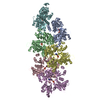
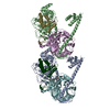
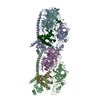
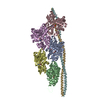
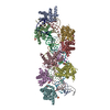
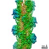
| Entry | Database: PDB / ID: 6u96 | ||||||||||||||||||||||||||||||
|---|---|---|---|---|---|---|---|---|---|---|---|---|---|---|---|---|---|---|---|---|---|---|---|---|---|---|---|---|---|---|---|
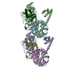
| Title | Actin phalloidin at BeFx state | ||||||||||||||||||||||||||||||
 Components Components |
| ||||||||||||||||||||||||||||||
 Keywords Keywords | STRUCTURAL PROTEIN / Actin / phalloidin / beryllium fluoride | ||||||||||||||||||||||||||||||
| Function / homology |  Function and homology information Function and homology informationcytoskeletal motor activator activity / myosin heavy chain binding / tropomyosin binding / actin filament bundle / troponin I binding / filamentous actin / mesenchyme migration / skeletal muscle myofibril / actin filament bundle assembly / striated muscle thin filament ...cytoskeletal motor activator activity / myosin heavy chain binding / tropomyosin binding / actin filament bundle / troponin I binding / filamentous actin / mesenchyme migration / skeletal muscle myofibril / actin filament bundle assembly / striated muscle thin filament / skeletal muscle thin filament assembly / actin monomer binding / skeletal muscle fiber development / stress fiber / titin binding / actin filament polymerization / filopodium / actin filament / Hydrolases; Acting on acid anhydrides; Acting on acid anhydrides to facilitate cellular and subcellular movement / calcium-dependent protein binding / lamellipodium / cell body / protein domain specific binding / hydrolase activity / calcium ion binding / positive regulation of gene expression / magnesium ion binding / ATP binding / identical protein binding / cytoplasm Similarity search - Function | ||||||||||||||||||||||||||||||
| Biological species |  synthetic construct (others) | ||||||||||||||||||||||||||||||
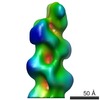
| Method | ELECTRON MICROSCOPY / helical reconstruction / cryo EM / Resolution: 3.8 Å | ||||||||||||||||||||||||||||||
 Authors Authors | Das, S. / Ge, P. / Durer, Z.A.O. / Grintsevich, E.E. / Zhou, Z.H. / Reisler, E. | ||||||||||||||||||||||||||||||
| Funding support |  United States, 4items United States, 4items
| ||||||||||||||||||||||||||||||
 Citation Citation | Journal: Structure / Year: 2020 Title: D-loop Dynamics and Near-Atomic-Resolution Cryo-EM Structure of Phalloidin-Bound F-Actin. Authors: Sanchaita Das / Peng Ge / Zeynep A Oztug Durer / Elena E Grintsevich / Z Hong Zhou / Emil Reisler / Abstract: Detailed molecular information on G-actin assembly into filaments (F-actin), and their structure, dynamics, and interactions, is essential for understanding their cellular functions. Previous studies ...Detailed molecular information on G-actin assembly into filaments (F-actin), and their structure, dynamics, and interactions, is essential for understanding their cellular functions. Previous studies indicate that a flexible DNase I binding loop (D-loop, residues 40-50) plays a major role in actin's conformational dynamics. Phalloidin, a "gold standard" for actin filament staining, stabilizes them and affects the D-loop. Using disulfide crosslinking in yeast actin D-loop mutant Q41C/V45C, light-scattering measurements, and cryoelectron microscopy reconstructions, we probed the constraints of D-loop dynamics and its contribution to F-actin formation/stability. Our data support a model of residues 41-45 distances that facilitate G- to F-actin transition. We report also a 3.3-Å resolution structure of phalloidin-bound F-actin in the ADP-Pi-like (ADP-BeFx) state. This shows the phalloidin-binding site on F-actin and how the relative movement between its two protofilaments is restricted by it. Together, our results provide molecular details of F-actin structure and D-loop dynamics. | ||||||||||||||||||||||||||||||
| History |
|
- Structure visualization
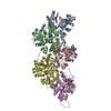
| Movie |
Movie viewer |
|---|---|
| Structure viewer | Molecule: MolmilJmol/JSmol |
- Downloads & links
-Download
| PDBx/mmCIF format | 6u96.cif.gz | 335.2 KB | Display | PDBx/mmCIF format |
|---|---|---|---|---|
| PDB format | pdb6u96.ent.gz | 274.5 KB | Display | PDB format |
| PDBx/mmJSON format | 6u96.json.gz | Tree view | PDBx/mmJSON format | |
| Others |  Other downloads Other downloads |
-Validation report
| Arichive directory | https://data.pdbj.org/pub/pdb/validation_reports/u9/6u96ftp://data.pdbj.org/pub/pdb/validation_reports/u9/6u96 | HTTPS FTP |
|---|
-Related structure data
| Related structure data | 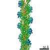 20694MC M: map data used to model this data C: citing same article ( |
|---|---|
| Similar structure data |

-Links
 PDBj
PDBj


- Assembly
| Deposited unit | 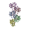
|
|---|---|
| 1 |
|
-Components
| #1: Protein | Mass: 42109.973 Da / Num. of mol.: 5 / Source method: isolated from a natural source / Details: ACTA1, ACTA / Source: (natural) #2: Protein/peptide | Mass: 808.899 Da / Num. of mol.: 5 / Source method: obtained synthetically / Source: (synth.) synthetic construct (others) #3: Chemical | ChemComp-ADP /   Mass: 427.201 Da / Num. of mol.: 5 / Source method: obtained synthetically / Formula: C10H15N5O10P2 / Feature type: SUBJECT OF INVESTIGATION / Comment: ADP, energy-carrying molecule*YM Mass: 427.201 Da / Num. of mol.: 5 / Source method: obtained synthetically / Formula: C10H15N5O10P2 / Feature type: SUBJECT OF INVESTIGATION / Comment: ADP, energy-carrying molecule*YMHas ligand of interest | Y | Has protein modification | Y | |
|---|
-Experimental details
-Experiment
| Experiment | Method: ELECTRON MICROSCOPY |
|---|---|
| EM experiment | Aggregation state: HELICAL ARRAY / 3D reconstruction method: helical reconstruction |
- Sample preparation
| Component | Name: Actin phalloidin, BeFx state / Type: ORGANELLE OR CELLULAR COMPONENT / Entity ID: #1 / Source: NATURAL | ||||||||||||||||||||
|---|---|---|---|---|---|---|---|---|---|---|---|---|---|---|---|---|---|---|---|---|---|
| Molecular weight | Experimental value: NO | ||||||||||||||||||||
| Source (natural) | Organism: | ||||||||||||||||||||
| Buffer solution | pH: 7.4 Details: pH7.4, 10mM Tris, 50mM KCl, 1 mM MgC2, 0.2mM CaCl2, 1mM ATP, 1mM DTT, 0.2mM EGTA, 0.2mM BeCl2, 5mM NaF | ||||||||||||||||||||
| Buffer component |
| ||||||||||||||||||||
| Specimen | Embedding applied: NO / Shadowing applied: NO / Staining applied: NO / Vitrification applied: YES | ||||||||||||||||||||
| Specimen support | Details: unspecified | ||||||||||||||||||||
| Vitrification | Instrument: FEI VITROBOT MARK IV / Cryogen name: ETHANE / Humidity: 100 % / Chamber temperature: 295 K |
- Electron microscopy imaging
| Experimental equipment |  Model: Titan Krios / Image courtesy: FEI Company |
|---|---|
| Microscopy | Model: FEI TITAN KRIOS |
| Electron gun | Electron source:  FIELD EMISSION GUN / Accelerating voltage: 300 kV / Illumination mode: FLOOD BEAM FIELD EMISSION GUN / Accelerating voltage: 300 kV / Illumination mode: FLOOD BEAM |
| Electron lens | Mode: BRIGHT FIELD / Nominal magnification: 130000 X / Nominal defocus max: 3000 nm / Nominal defocus min: 3000 nm / Calibrated defocus min: 2200 nm / Calibrated defocus max: 4200 nm / Cs: 2.7 mm / C2 aperture diameter: 70 µm / Alignment procedure: COMA FREE |
| Specimen holder | Cryogen: NITROGEN / Specimen holder model: FEI TITAN KRIOS AUTOGRID HOLDER / Temperature (max): 81 K / Temperature (min): 80 K |
| Image recording | Average exposure time: 8 sec. / Electron dose: 80 e/Å2 / Detector mode: COUNTING / Film or detector model: GATAN K2 QUANTUM (4k x 4k) / Num. of grids imaged: 1 / Num. of real images: 5874 |
| EM imaging optics | Energyfilter name: GIF Quantum LS |
| Image scans | Movie frames/image: 32 / Used frames/image: 3-12 |
- Processing
| Software | Name: PHENIX / Version: 1.13_2998: / Classification: refinement | ||||||||||||||||||||||||||||
|---|---|---|---|---|---|---|---|---|---|---|---|---|---|---|---|---|---|---|---|---|---|---|---|---|---|---|---|---|---|
| EM software |
| ||||||||||||||||||||||||||||
| CTF correction | Type: PHASE FLIPPING AND AMPLITUDE CORRECTION | ||||||||||||||||||||||||||||
| Helical symmerty | Angular rotation/subunit: -166.967625 ° / Axial rise/subunit: 29.508839 Å / Axial symmetry: C1 | ||||||||||||||||||||||||||||
| Particle selection | Num. of particles selected: 214555 | ||||||||||||||||||||||||||||
| 3D reconstruction | Resolution: 3.8 Å / Resolution method: FSC 0.143 CUT-OFF / Num. of particles: 181626 / Symmetry type: HELICAL | ||||||||||||||||||||||||||||
| Atomic model building | Protocol: AB INITIO MODEL / Space: REAL |
