 Movie
Movie Controller

[English] 日本語
 Yorodumi
- PDB-6qnu: Human Adenovirus type 3 fiber knob in complex with two copies of ... -
+ Open data

- Basic information
| Entry | Database: PDB / ID: 6qnu | ||||||
|---|---|---|---|---|---|---|---|
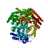
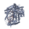
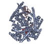
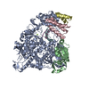
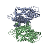
| Title | Human Adenovirus type 3 fiber knob in complex with two copies of Desmoglein-2 | ||||||
 Components Components |
| ||||||
 Keywords Keywords | VIRAL PROTEIN / virus receptor / adenovirus / vector | ||||||
| Function / homology |  Function and homology information Function and homology informationPurkinje myocyte development / positive regulation of protein localization to cell-cell junction / bundle of His cell-Purkinje myocyte adhesion involved in cell communication / cell adhesive protein binding involved in bundle of His cell-Purkinje myocyte communication / desmosome organization / negative regulation of endothelial cell differentiation / Keratinization / negative regulation of inflammatory response to wounding / desmosome / mesenchymal to epithelial transition ...Purkinje myocyte development / positive regulation of protein localization to cell-cell junction / bundle of His cell-Purkinje myocyte adhesion involved in cell communication / cell adhesive protein binding involved in bundle of His cell-Purkinje myocyte communication / desmosome organization / negative regulation of endothelial cell differentiation / Keratinization / negative regulation of inflammatory response to wounding / desmosome / mesenchymal to epithelial transition / Formation of the cornified envelope / cornified envelope / regulation of ventricular cardiac muscle cell action potential / Apoptotic cleavage of cell adhesion proteins / adhesion receptor-mediated virion attachment to host cell / negative regulation of epithelial to mesenchymal transition / positive regulation of sprouting angiogenesis / homophilic cell-cell adhesion / positive regulation of stem cell population maintenance / regulation of heart rate by cardiac conduction / RHOG GTPase cycle / intercalated disc / lateral plasma membrane / RAC3 GTPase cycle / maternal process involved in female pregnancy / RAC2 GTPase cycle / response to progesterone / cell adhesion molecule binding / positive regulation of cell adhesion / stem cell proliferation / cell-cell adhesion / cell-cell junction / viral capsid / cell junction / cell adhesion / apical plasma membrane / calcium ion binding / symbiont entry into host cell / negative regulation of apoptotic process / host cell nucleus / cell surface / extracellular exosome / plasma membrane / cytoplasm Similarity search - Function | ||||||
| Biological species |  Human adenovirus B serotype 3 Human adenovirus B serotype 3 Homo sapiens (human) Homo sapiens (human) | ||||||
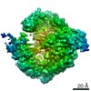
| Method | ELECTRON MICROSCOPY / single particle reconstruction / cryo EM / Resolution: 3.8 Å | ||||||
 Authors Authors | Effantin, G. | ||||||
| Funding support |  France, 1items France, 1items
| ||||||
 Citation Citation | Journal: Nat Commun / Year: 2019 Title: CryoEM structure of adenovirus type 3 fibre with desmoglein 2 shows an unusual mode of receptor engagement. Authors: Emilie Vassal-Stermann / Gregory Effantin / Chloe Zubieta / Wim Burmeister / Frédéric Iseni / Hongjie Wang / André Lieber / Guy Schoehn / Pascal Fender /  Abstract: Attachment of human adenovirus (HAd) to the host cell is a critical step of infection. Initial attachment occurs via the adenoviral fibre knob protein and a cellular receptor. Here we report the cryo- ...Attachment of human adenovirus (HAd) to the host cell is a critical step of infection. Initial attachment occurs via the adenoviral fibre knob protein and a cellular receptor. Here we report the cryo-electron microscopy (cryo-EM) structure of a <100 kDa non-symmetrical complex comprising the trimeric HAd type 3 fibre knob (HAd3K) and human desmoglein 2 (DSG2). The structure reveals a unique stoichiometry of 1:1 and 2:1 (DSG2: knob trimer) not previously observed for other HAd-receptor complexes. We demonstrate that mutating Asp261 in the fibre knob is sufficient to totally abolish receptor binding. These data shed new light on adenovirus infection strategies and provide insights for adenoviral vector development and structure-based design. | ||||||
| History |
|
- Structure visualization
| Movie |
Movie viewer |
|---|---|
| Structure viewer | Molecule: MolmilJmol/JSmol |
- Downloads & links
-Download
| PDBx/mmCIF format | 6qnu.cif.gz | 185.1 KB | Display | PDBx/mmCIF format |
|---|---|---|---|---|
| PDB format | pdb6qnu.ent.gz | 146.6 KB | Display | PDB format |
| PDBx/mmJSON format | 6qnu.json.gz | Tree view | PDBx/mmJSON format | |
| Others |  Other downloads Other downloads |
-Validation report
| Arichive directory | https://data.pdbj.org/pub/pdb/validation_reports/qn/6qnuftp://data.pdbj.org/pub/pdb/validation_reports/qn/6qnu | HTTPS FTP |
|---|
-Related structure data
| Related structure data | 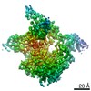 4609MC 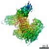 4608C  6qntC C: citing same article ( M: map data used to model this data |
|---|---|
| Similar structure data |


-Links
 PDBj
PDBj


- Assembly
| Deposited unit | 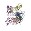
|
|---|---|
| 1 |
|
-Components
| #1: Protein | Mass: 21025.748 Da / Num. of mol.: 3 Source method: isolated from a genetically manipulated source Source: (gene. exp.) Human adenovirus B serotype 3 / Gene: L5 / Production host:  #2: Protein | Mass: 25055.029 Da / Num. of mol.: 2 Source method: isolated from a genetically manipulated source Source: (gene. exp.) Homo sapiens (human) / Gene: DSG2, CDHF5 / Production host: |
|---|
-Experimental details
-Experiment
| Experiment | Method: ELECTRON MICROSCOPY |
|---|---|
| EM experiment | Aggregation state: PARTICLE / 3D reconstruction method: single particle reconstruction |
- Sample preparation
| Component |
| ||||||||||||||||||||||||
|---|---|---|---|---|---|---|---|---|---|---|---|---|---|---|---|---|---|---|---|---|---|---|---|---|---|
| Molecular weight | Value: 0.116 MDa / Experimental value: YES | ||||||||||||||||||||||||
| Source (natural) |
| ||||||||||||||||||||||||
| Source (recombinant) |
| ||||||||||||||||||||||||
| Buffer solution | pH: 8 | ||||||||||||||||||||||||
| Specimen | Embedding applied: NO / Shadowing applied: NO / Staining applied: NO / Vitrification applied: YES | ||||||||||||||||||||||||
| Vitrification | Cryogen name: ETHANE |
- Electron microscopy imaging
| Experimental equipment |  Model: Titan Krios / Image courtesy: FEI Company |
|---|---|
| Microscopy | Model: FEI TITAN KRIOS |
| Electron gun | Electron source:  FIELD EMISSION GUN / Accelerating voltage: 300 kV / Illumination mode: FLOOD BEAM FIELD EMISSION GUN / Accelerating voltage: 300 kV / Illumination mode: FLOOD BEAM |
| Electron lens | Mode: BRIGHT FIELD |
| Image recording | Electron dose: 35 e/Å2 / Film or detector model: GATAN K2 SUMMIT (4k x 4k) |
| EM imaging optics | Phase plate: VOLTA PHASE PLATE |
- Processing
| CTF correction | Type: PHASE FLIPPING AND AMPLITUDE CORRECTION |
|---|---|
| Symmetry | Point symmetry: C1 (asymmetric) |
| 3D reconstruction | Resolution: 3.8 Å / Resolution method: FSC 0.143 CUT-OFF / Num. of particles: 78925 / Symmetry type: POINT |
