 Movie
Movie Controller

[English] 日本語
 Yorodumi
- PDB-6ppr: Cryo-EM structure of AdnA(D934A)-AdnB(D1014A) in complex with AMP... -
+ Open data

- Basic information
| Entry | Database: PDB / ID: 6ppr | ||||||
|---|---|---|---|---|---|---|---|
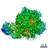
| Title | Cryo-EM structure of AdnA(D934A)-AdnB(D1014A) in complex with AMPPNP and DNA | ||||||
 Components Components |
| ||||||
 Keywords Keywords | DNA BINDING PROTEIN/DNA / DNA / DNA BINDING PROTEIN / DNA BINDING PROTEIN-DNA complex | ||||||
| Function / homology |  Function and homology information Function and homology informationDNA helicase complex / DNA 3'-5' helicase / recombinational repair / exonuclease activity / 3'-5' DNA helicase activity / DNA helicase activity / DNA helicase / hydrolase activity / DNA repair / DNA binding ...DNA helicase complex / DNA 3'-5' helicase / recombinational repair / exonuclease activity / 3'-5' DNA helicase activity / DNA helicase activity / DNA helicase / hydrolase activity / DNA repair / DNA binding / ATP binding / cytosol Similarity search - Function | ||||||
| Biological species |  Mycobacterium smegmatis (bacteria)Mycolicibacterium smegmatis (bacteria) Mycobacterium smegmatis (bacteria)Mycolicibacterium smegmatis (bacteria) | ||||||
| Method | ELECTRON MICROSCOPY / single particle reconstruction / cryo EM / Resolution: 3.5 Å | ||||||
 Authors Authors | Jia, N. / Unciuleac, M. / Shuman, S. / Patel, D.J. | ||||||
 Citation Citation | Journal: Proc Natl Acad Sci U S A / Year: 2019 Title: Structures and single-molecule analysis of bacterial motor nuclease AdnAB illuminate the mechanism of DNA double-strand break resection. Authors: Ning Jia / Mihaela C Unciuleac / Chaoyou Xue / Eric C Greene / Dinshaw J Patel / Stewart Shuman /  Abstract: Mycobacterial AdnAB is a heterodimeric helicase-nuclease that initiates homologous recombination by resecting DNA double-strand breaks (DSBs). The AdnA and AdnB subunits are each composed of an N- ...Mycobacterial AdnAB is a heterodimeric helicase-nuclease that initiates homologous recombination by resecting DNA double-strand breaks (DSBs). The AdnA and AdnB subunits are each composed of an N-terminal motor domain and a C-terminal nuclease domain. Here we report cryoelectron microscopy (cryo-EM) structures of AdnAB in three functional states: in the absence of DNA and in complex with forked duplex DNAs before and after cleavage of the 5' single-strand DNA (ssDNA) tail by the AdnA nuclease. The structures reveal the path of the 5' ssDNA through the AdnA nuclease domain and the mechanism of 5' strand cleavage; the path of the 3' tracking strand through the AdnB motor and the DNA contacts that couple ATP hydrolysis to mechanical work; the position of the AdnA iron-sulfur cluster subdomain at the Y junction and its likely role in maintaining the split trajectories of the unwound 5' and 3' strands. Single-molecule DNA curtain analysis of DSB resection reveals that AdnAB is highly processive but prone to spontaneous pausing at random sites on duplex DNA. A striking property of AdnAB is that the velocity of DSB resection slows after the enzyme experiences a spontaneous pause. Our results highlight shared as well as distinctive properties of AdnAB vis-à-vis the RecBCD and AddAB clades of bacterial DSB-resecting motor nucleases. | ||||||
| History |
|
- Structure visualization
| Movie |
Movie viewer |
|---|---|
| Structure viewer | Molecule: MolmilJmol/JSmol |
- Downloads & links
-Download
| PDBx/mmCIF format | 6ppr.cif.gz | 299.8 KB | Display | PDBx/mmCIF format |
|---|---|---|---|---|
| PDB format | pdb6ppr.ent.gz | 222.8 KB | Display | PDB format |
| PDBx/mmJSON format | 6ppr.json.gz | Tree view | PDBx/mmJSON format | |
| Others |  Other downloads Other downloads |
-Validation report
| Summary document | 6ppr_validation.pdf.gz | 884.4 KB | Display | wwPDB validaton report |
|---|---|---|---|---|
| Full document | 6ppr_full_validation.pdf.gz | 906.4 KB | Display | |
| Data in XML | 6ppr_validation.xml.gz | 51.8 KB | Display | |
| Data in CIF | 6ppr_validation.cif.gz | 80 KB | Display | |
| Arichive directory | https://data.pdbj.org/pub/pdb/validation_reports/pp/6pprftp://data.pdbj.org/pub/pdb/validation_reports/pp/6ppr | HTTPS FTP |
-Related structure data
| Related structure data |  20446MC  6ppjC  6ppuC C: citing same article ( M: map data used to model this data |
|---|---|
| Similar structure data |


-Links
 PDBj
PDBj


- Assembly
| Deposited unit | 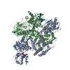
|
|---|---|
| 1 |
|
-Components
| #1: Protein | Mass: 118084.531 Da / Num. of mol.: 1 Source method: isolated from a genetically manipulated source Source: (gene. exp.) Mycobacterium smegmatis (bacteria) / Gene: pcrA_2, ERS451418_01974 / Production host: References: UniProt: A0A0D6HIW1, UniProt: I7FZ56*PLUS, DNA helicase |
|---|---|
| #2: Protein | Mass: 110899.562 Da / Num. of mol.: 1 Source method: isolated from a genetically manipulated source Source: (gene. exp.) Mycobacterium smegmatis (bacteria) / Gene: pcrA_1, ERS451418_01973 / Production host: References: UniProt: A0A0D6HKQ2, UniProt: A0QTR9*PLUS, DNA helicase |
| #3: DNA chain | Mass: 21477.703 Da / Num. of mol.: 1 / Source method: obtained synthetically / Source: (synth.) Mycolicibacterium smegmatis (bacteria) |
| #4: Chemical | ChemComp-ANP /   Mass: 506.196 Da / Num. of mol.: 1 / Source method: obtained synthetically / Formula: C10H17N6O12P3 / Comment: AMP-PNP, energy-carrying molecule analogue*YM Mass: 506.196 Da / Num. of mol.: 1 / Source method: obtained synthetically / Formula: C10H17N6O12P3 / Comment: AMP-PNP, energy-carrying molecule analogue*YM |
| #5: Chemical | ChemComp-SF4 /   Mass: 351.640 Da / Num. of mol.: 1 / Source method: obtained synthetically / Formula: Fe4S4 Mass: 351.640 Da / Num. of mol.: 1 / Source method: obtained synthetically / Formula: Fe4S4 |
-Experimental details
-Experiment
| Experiment | Method: ELECTRON MICROSCOPY |
|---|---|
| EM experiment | Aggregation state: PARTICLE / 3D reconstruction method: single particle reconstruction |
- Sample preparation
| Component |
| ||||||||||||||||||||||||||||||
|---|---|---|---|---|---|---|---|---|---|---|---|---|---|---|---|---|---|---|---|---|---|---|---|---|---|---|---|---|---|---|---|
| Molecular weight | Value: 0.2 MDa / Experimental value: YES | ||||||||||||||||||||||||||||||
| Source (natural) |
| ||||||||||||||||||||||||||||||
| Source (recombinant) |
| ||||||||||||||||||||||||||||||
| Buffer solution | pH: 7.5 / Details: 20 mM Tris-HCl, pH 7.5, 150 mM NaCl | ||||||||||||||||||||||||||||||
| Buffer component | Formula: Tris | ||||||||||||||||||||||||||||||
| Specimen | Conc.: 1.5 mg/ml / Embedding applied: NO / Shadowing applied: NO / Staining applied: NO / Vitrification applied: YES | ||||||||||||||||||||||||||||||
| Vitrification | Instrument: FEI VITROBOT MARK IV / Cryogen name: ETHANE / Humidity: 100 % |
- Electron microscopy imaging
| Experimental equipment |  Model: Titan Krios / Image courtesy: FEI Company |
|---|---|
| Microscopy | Model: FEI TITAN KRIOS |
| Electron gun | Electron source:  FIELD EMISSION GUN / Accelerating voltage: 300 kV / Illumination mode: FLOOD BEAM FIELD EMISSION GUN / Accelerating voltage: 300 kV / Illumination mode: FLOOD BEAM |
| Electron lens | Mode: BRIGHT FIELD |
| Image recording | Electron dose: 2.16 e/Å2 / Film or detector model: GATAN K2 SUMMIT (4k x 4k) |
- Processing
| EM software | Name: RELION / Version: 2.1 / Category: 3D reconstruction |
|---|---|
| CTF correction | Type: PHASE FLIPPING AND AMPLITUDE CORRECTION |
| 3D reconstruction | Resolution: 3.5 Å / Resolution method: FSC 0.143 CUT-OFF / Num. of particles: 61579 / Symmetry type: POINT |
