host cellular component / Synthesis And Processing Of GAG, GAGPOL Polyproteins / host cell nuclear membrane / Integration of viral DNA into host genomic DNA / Autointegration results in viral DNA circles / Minus-strand DNA synthesis / Plus-strand DNA synthesis / 2-LTR circle formation / Uncoating of the HIV Virion / viral budding via host ESCRT complex ...host cellular component / Synthesis And Processing Of GAG, GAGPOL Polyproteins / host cell nuclear membrane / Integration of viral DNA into host genomic DNA / Autointegration results in viral DNA circles / Minus-strand DNA synthesis / Plus-strand DNA synthesis / 2-LTR circle formation / Uncoating of the HIV Virion / viral budding via host ESCRT complex / Vpr-mediated nuclear import of PICs / Early Phase of HIV Life Cycle / Integration of provirus / APOBEC3G mediated resistance to HIV-1 infection / Binding and entry of HIV virion / Membrane binding and targetting of GAG proteins / viral process / Assembly Of The HIV Virion / Budding and maturation of HIV virion / host multivesicular body / viral capsid / viral nucleocapsid / host cell plasma membrane / structural molecule activity / virion membrane / RNA binding / zinc ion binding / membrane 類似検索 - 分子機能
Retrovirus capsid C-terminal domain / Gag protein p6 / Gag protein p6 / Non-ribosomal Peptide Synthetase Peptidyl Carrier Protein; Chain A / gag protein p24 N-terminal domain / Immunodeficiency lentiviral matrix, N-terminal / gag gene protein p17 (matrix protein) / Retroviral nucleocapsid Gag protein p24, C-terminal domain / Gag protein p24 C-terminal domain / Matrix protein, lentiviral and alpha-retroviral, N-terminal ...Retrovirus capsid C-terminal domain / Gag protein p6 / Gag protein p6 / Non-ribosomal Peptide Synthetase Peptidyl Carrier Protein; Chain A / gag protein p24 N-terminal domain / Immunodeficiency lentiviral matrix, N-terminal / gag gene protein p17 (matrix protein) / Retroviral nucleocapsid Gag protein p24, C-terminal domain / Gag protein p24 C-terminal domain / Matrix protein, lentiviral and alpha-retroviral, N-terminal / Retrovirus capsid, C-terminal / Retroviral matrix protein / Retrovirus capsid, N-terminal / zinc finger / Zinc knuckle / Zinc finger, CCHC-type superfamily / Zinc finger, CCHC-type / Zinc finger CCHC-type profile. / Orthogonal Bundle / Mainly Alpha 類似検索 - ドメイン・相同性
National Institutes of Health/National Human Genome Research Institute (NIH/NHGRI)
5 P50 GM082545-10
米国
National Institutes of Health/National Institute of General Medical Sciences (NIH/NIGMS)
R01 GM066087
米国
National Institutes of Health/National Institute of General Medical Sciences (NIH/NIGMS)
P50 GM082545
米国
National Institutes of Health/National Institute of General Medical Sciences (NIH/NIGMS)
R01 GM12850
米国
引用
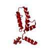
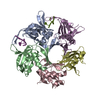
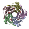
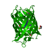
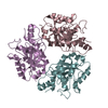
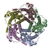
ジャーナル: Proc Natl Acad Sci U S A / 年: 2018 タイトル: MicroED structures of HIV-1 Gag CTD-SP1 reveal binding interactions with the maturation inhibitor bevirimat. 著者: Michael D Purdy / Dan Shi / Jakub Chrustowicz / Johan Hattne / Tamir Gonen / Mark Yeager / 要旨: HIV-1 protease (PR) cleavage of the Gag polyprotein triggers the assembly of mature, infectious particles. Final cleavage of Gag occurs at the junction helix between the capsid protein CA and the SP1 ...HIV-1 protease (PR) cleavage of the Gag polyprotein triggers the assembly of mature, infectious particles. Final cleavage of Gag occurs at the junction helix between the capsid protein CA and the SP1 spacer peptide. Here we used MicroED to delineate the binding interactions of the maturation inhibitor bevirimat (BVM) using very thin frozen-hydrated, 3D microcrystals of a CTD-SP1 Gag construct with and without bound BVM. The 2.9-Å MicroED structure revealed that a single BVM molecule stabilizes the six-helix bundle via both electrostatic interactions with the dimethylsuccinyl moiety and hydrophobic interactions with the pentacyclic triterpenoid ring. These results provide insight into the mechanism of action of BVM and related maturation inhibitors that will inform further drug discovery efforts. This study also demonstrates the capabilities of MicroED for structure-based drug design.
A: CTD-SP1 fragment of HIV-1 Gag B: CTD-SP1 fragment of HIV-1 Gag C: CTD-SP1 fragment of HIV-1 Gag D: CTD-SP1 fragment of HIV-1 Gag E: CTD-SP1 fragment of HIV-1 Gag F: CTD-SP1 fragment of HIV-1 Gag
平均露光時間: 8 sec. / 電子線照射量: 0.05 e/Å2 フィルム・検出器のモデル: TVIPS TEMCAM-F416 (4k x 4k) 撮影したグリッド数: 6 詳細: Data from 6 crystals was merged for structure determination.
 ムービー
ムービー コントローラー


 データを開く
 基本情報
基本情報 要素
要素 キーワード
キーワード 機能・相同性情報
 Human immunodeficiency virus 1 (ヒト免疫不全ウイルス)
 分子置換 / クライオ電子顕微鏡法 / 解像度: 3 Å
  データ登録者
 米国, 4件
  引用
引用 構造の表示
構造の表示 ダウンロードとリンク
 その他のダウンロード


 PDBj
PDBj


 集合体
集合体

 試料調製
試料調製
 解析
解析