 Movie
Movie Controller

[English] 日本語
 Yorodumi
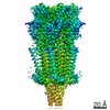
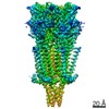
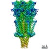
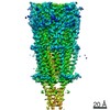
- PDB-6his: Mouse serotonin 5-HT3 receptor, tropisetron-bound, T conformation -
+ Open data

- Basic information
| Entry | Database: PDB / ID: 6his | ||||||||||||
|---|---|---|---|---|---|---|---|---|---|---|---|---|---|
| Title | Mouse serotonin 5-HT3 receptor, tropisetron-bound, T conformation | ||||||||||||
 Components Components | 5-hydroxytryptamine receptor 3A | ||||||||||||
 Keywords Keywords | MEMBRANE PROTEIN / Ion channel / serotonin receptor / pentameric ligand-gated channel | ||||||||||||
| Function / homology |  Function and homology information Function and homology informationNeurotransmitter receptors and postsynaptic signal transmission / serotonin-gated cation-selective signaling pathway / serotonin-activated cation-selective channel complex / serotonin-gated monoatomic cation channel activity / serotonin receptor signaling pathway / serotonin binding / transmembrane transporter complex / excitatory extracellular ligand-gated monoatomic ion channel activity / : / cleavage furrow ...Neurotransmitter receptors and postsynaptic signal transmission / serotonin-gated cation-selective signaling pathway / serotonin-activated cation-selective channel complex / serotonin-gated monoatomic cation channel activity / serotonin receptor signaling pathway / serotonin binding / transmembrane transporter complex / excitatory extracellular ligand-gated monoatomic ion channel activity / : / cleavage furrow / ligand-gated monoatomic ion channel activity involved in regulation of presynaptic membrane potential / transmitter-gated monoatomic ion channel activity involved in regulation of postsynaptic membrane potential / regulation of membrane potential / presynaptic membrane / monoatomic ion transmembrane transport / chemical synaptic transmission / postsynaptic membrane / neuron projection / axon / neuronal cell body / synapse / glutamatergic synapse / identical protein binding / plasma membrane Similarity search - Function | ||||||||||||
| Biological species |  | ||||||||||||
| Method | ELECTRON MICROSCOPY / single particle reconstruction / cryo EM / Resolution: 4.5 Å | ||||||||||||
 Authors Authors | Polovinkin, L. / Neumann, E. / Schoehn, G. / Nury, H. | ||||||||||||
 Citation Citation | Journal: Nature / Year: 2018 Title: Conformational transitions of the serotonin 5-HT receptor. Authors: Lucie Polovinkin / Ghérici Hassaine / Jonathan Perot / Emmanuelle Neumann / Anders A Jensen / Solène N Lefebvre / Pierre-Jean Corringer / Jacques Neyton / Christophe Chipot / Francois ...Authors: Lucie Polovinkin / Ghérici Hassaine / Jonathan Perot / Emmanuelle Neumann / Anders A Jensen / Solène N Lefebvre / Pierre-Jean Corringer / Jacques Neyton / Christophe Chipot / Francois Dehez / Guy Schoehn / Hugues Nury /    Abstract: The serotonin 5-HT receptor is a pentameric ligand-gated ion channel (pLGIC). It belongs to a large family of receptors that function as allosteric signal transducers across the plasma membrane; upon ...The serotonin 5-HT receptor is a pentameric ligand-gated ion channel (pLGIC). It belongs to a large family of receptors that function as allosteric signal transducers across the plasma membrane; upon binding of neurotransmitter molecules to extracellular sites, the receptors undergo complex conformational transitions that result in transient opening of a pore permeable to ions. 5-HT receptors are therapeutic targets for emesis and nausea, irritable bowel syndrome and depression. In spite of several reported pLGIC structures, no clear unifying view has emerged on the conformational transitions involved in channel gating. Here we report four cryo-electron microscopy structures of the full-length mouse 5-HT receptor in complex with the anti-emetic drug tropisetron, with serotonin, and with serotonin and a positive allosteric modulator, at resolutions ranging from 3.2 Å to 4.5 Å. The tropisetron-bound structure resembles those obtained with an inhibitory nanobody or without ligand. The other structures include an 'open' state and two ligand-bound states. We present computational insights into the dynamics of the structures, their pore hydration and free-energy profiles, and characterize movements at the gate level and cation accessibility in the pore. Together, these data deepen our understanding of the gating mechanism of pLGICs and capture ligand binding in unprecedented detail. | ||||||||||||
| History |
|
- Structure visualization
| Movie |
Movie viewer |
|---|---|
| Structure viewer | Molecule: MolmilJmol/JSmol |
- Downloads & links
-Download
| PDBx/mmCIF format | 6his.cif.gz | 362.8 KB | Display | PDBx/mmCIF format |
|---|---|---|---|---|
| PDB format | pdb6his.ent.gz | 298.5 KB | Display | PDB format |
| PDBx/mmJSON format | 6his.json.gz | Tree view | PDBx/mmJSON format | |
| Others |  Other downloads Other downloads |
-Validation report
| Arichive directory | https://data.pdbj.org/pub/pdb/validation_reports/hi/6hisftp://data.pdbj.org/pub/pdb/validation_reports/hi/6his | HTTPS FTP |
|---|
-Related structure data
| Related structure data | 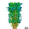 0228MC  0225C 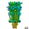 0226C 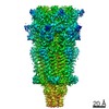 0227C  6hinC  6hioC  6hiqC C: citing same article ( M: map data used to model this data |
|---|---|
| Similar structure data |


-Links
 PDBj
PDBj


- Assembly
| Deposited unit | 
|
|---|---|
| 1 |
|
-Components
| #1: Protein | Mass: 51949.055 Da / Num. of mol.: 5 Source method: isolated from a genetically manipulated source Source: (gene. exp.)  Homo sapiens (human) / References: UniProt: P23979 Homo sapiens (human) / References: UniProt: P23979#2: Polysaccharide | 2-acetamido-2-deoxy-beta-D-glucopyranose-(1-4)-2-acetamido-2-deoxy-beta-D-glucopyranose Source method: isolated from a genetically manipulated source #3: Polysaccharide | beta-D-mannopyranose-(1-4)-2-acetamido-2-deoxy-beta-D-glucopyranose-(1-4)-2-acetamido-2-deoxy-beta- ...beta-D-mannopyranose-(1-4)-2-acetamido-2-deoxy-beta-D-glucopyranose-(1-4)-2-acetamido-2-deoxy-beta-D-glucopyranose Source method: isolated from a genetically manipulated source #4: Chemical | ChemComp-TKT / ( 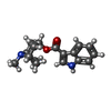  Mass: 284.353 Da / Num. of mol.: 5 / Source method: obtained synthetically / Formula: C17H20N2O2 / Comment: chemotherapy, antagonist*YM Mass: 284.353 Da / Num. of mol.: 5 / Source method: obtained synthetically / Formula: C17H20N2O2 / Comment: chemotherapy, antagonist*YMHas protein modification | Y | |
|---|
-Experimental details
-Experiment
| Experiment | Method: ELECTRON MICROSCOPY |
|---|---|
| EM experiment | Aggregation state: PARTICLE / 3D reconstruction method: single particle reconstruction |
- Sample preparation
| Component | Name: 5-HT3 receptor, serotonin-bound / Type: COMPLEX / Entity ID: #1 / Source: RECOMBINANT |
|---|---|
| Molecular weight | Experimental value: NO |
| Source (natural) | Organism: |
| Source (recombinant) | Organism: Homo sapiens (human) / Cell: HEK 293 |
| Buffer solution | pH: 7.4 |
| Specimen | Embedding applied: NO / Shadowing applied: NO / Staining applied: NO / Vitrification applied: YES |
| Vitrification | Cryogen name: ETHANE |
- Electron microscopy imaging
| Experimental equipment |  Model: Titan Krios / Image courtesy: FEI Company |
|---|---|
| Microscopy | Model: FEI TITAN KRIOS |
| Electron gun | Electron source:  FIELD EMISSION GUN / Accelerating voltage: 300 kV / Illumination mode: FLOOD BEAM FIELD EMISSION GUN / Accelerating voltage: 300 kV / Illumination mode: FLOOD BEAM |
| Electron lens | Mode: BRIGHT FIELD |
| Image recording | Electron dose: 80 e/Å2 / Film or detector model: GATAN K2 SUMMIT (4k x 4k) |
- Processing
| CTF correction | Type: PHASE FLIPPING AND AMPLITUDE CORRECTION |
|---|---|
| Symmetry | Point symmetry: C5 (5 fold cyclic) |
| 3D reconstruction | Resolution: 4.5 Å / Resolution method: FSC 0.143 CUT-OFF / Num. of particles: 43558 / Symmetry type: POINT |
