 Movie
Movie Controller


+ Open data

- Basic information
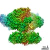
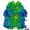
| Entry | Database: PDB / ID: 6h3i | |||||||||
|---|---|---|---|---|---|---|---|---|---|---|
| Title | Structural snapshots of the Type 9 protein translocon | |||||||||
 Components Components |
| |||||||||
 Keywords Keywords | PROTEIN TRANSPORT / Type 9 Secretion System Type IX Secretion System T9S folded protein secretion outer membrane protein | |||||||||
| Function / homology |  Function and homology information Function and homology information | |||||||||
| Biological species |  Flavobacterium johnsoniae (bacteria) Flavobacterium johnsoniae (bacteria) | |||||||||
| Method | ELECTRON MICROSCOPY / single particle reconstruction / cryo EM / Resolution: 3.5 Å | |||||||||
 Authors Authors | Deme, J.C. / Lea, S.M. | |||||||||
| Funding support |  United Kingdom, 2items United Kingdom, 2items
| |||||||||
 Citation Citation | Journal: Nature / Year: 2018 Title: Type 9 secretion system structures reveal a new protein transport mechanism. Authors: Frédéric Lauber / Justin C Deme / Susan M Lea / Ben C Berks / Abstract: The type 9 secretion system (T9SS) is the protein export pathway of bacteria of the Gram-negative Fibrobacteres-Chlorobi-Bacteroidetes superphylum and is an essential determinant of pathogenicity in ...The type 9 secretion system (T9SS) is the protein export pathway of bacteria of the Gram-negative Fibrobacteres-Chlorobi-Bacteroidetes superphylum and is an essential determinant of pathogenicity in severe periodontal disease. The central element of the T9SS is a so-far uncharacterized protein-conducting translocon located in the bacterial outer membrane. Here, using cryo-electron microscopy, we provide structural evidence that the translocon is the T9SS protein SprA. SprA forms an extremely large (36-strand) single polypeptide transmembrane β-barrel. The barrel pore is capped on the extracellular end, but has a lateral opening to the external membrane surface. Structures of SprA bound to different components of the T9SS show that partner proteins control access to the lateral opening and to the periplasmic end of the pore. Our results identify a protein transporter with a distinctive architecture that uses an alternating access mechanism in which the two ends of the protein-conducting channel are open at different times. | |||||||||
| History |
|
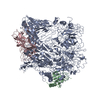
- Structure visualization
| Movie |
Movie viewer |
|---|---|
| Structure viewer | Molecule: MolmilJmol/JSmol |
- Downloads & links
-Download
| PDBx/mmCIF format | 6h3i.cif.gz | 473.8 KB | Display | PDBx/mmCIF format |
|---|---|---|---|---|
| PDB format | pdb6h3i.ent.gz | 371.3 KB | Display | PDB format |
| PDBx/mmJSON format | 6h3i.json.gz | Tree view | PDBx/mmJSON format | |
| Others |  Other downloads Other downloads |
-Validation report
| Arichive directory | https://data.pdbj.org/pub/pdb/validation_reports/h3/6h3iftp://data.pdbj.org/pub/pdb/validation_reports/h3/6h3i | HTTPS FTP |
|---|
-Related structure data
| Related structure data | 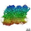 0133MC  0134C 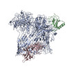 6h3jC M: map data used to model this data C: citing same article ( |
|---|---|
| Similar structure data |

-Links
 PDBj
PDBj


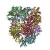
- Assembly
| Deposited unit | 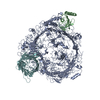
|
|---|---|
| 1 |
|
-Components
| #1: Protein | Mass: 270221.688 Da / Num. of mol.: 1 / Source method: isolated from a natural source Details: SprA from flavobacterium johnsoniae uniprot Q5I6C7 fjoh_1653 Source: (natural) Flavobacterium johnsoniae (bacteria) / References: UniProt: A0A1M5G5I4, UniProt: Q5I6C7*PLUS |
|---|---|
| #2: Protein | Mass: 19219.141 Da / Num. of mol.: 1 / Source method: isolated from a natural source / Source: (natural) Flavobacterium johnsoniae (bacteria) / Strain: ATCC 17061 / DSM 2064 / UW101 / References: UniProt: A5F9W9, peptidylprolyl isomerase |
| #3: Protein | Mass: 44210.043 Da / Num. of mol.: 1 / Source method: isolated from a natural source / Source: (natural) Flavobacterium johnsoniae (bacteria) / Strain: ATCC 17061 / DSM 2064 / UW101 / References: UniProt: A5FJM7 |
-Experimental details
-Experiment
| Experiment | Method: ELECTRON MICROSCOPY |
|---|---|
| EM experiment | Aggregation state: PARTICLE / 3D reconstruction method: single particle reconstruction |
- Sample preparation
| Component | Name: Complex of SprA, PPI and PorV / Type: COMPLEX / Entity ID: all / Source: NATURAL |
|---|---|
| Molecular weight | Value: 0.335 MDa / Experimental value: NO |
| Source (natural) | Organism: Flavobacterium johnsoniae (bacteria) |
| Buffer solution | pH: 7.5 |
| Specimen | Embedding applied: NO / Shadowing applied: NO / Staining applied: NO / Vitrification applied: YES |
| Vitrification | Cryogen name: ETHANE |
- Electron microscopy imaging
| Experimental equipment |  Model: Titan Krios / Image courtesy: FEI Company |
|---|---|
| Microscopy | Model: FEI TITAN KRIOS |
| Electron gun | Electron source:  FIELD EMISSION GUN / Accelerating voltage: 300 kV / Illumination mode: SPOT SCAN FIELD EMISSION GUN / Accelerating voltage: 300 kV / Illumination mode: SPOT SCAN |
| Electron lens | Mode: BRIGHT FIELD |
| Image recording | Electron dose: 52 e/Å2 / Detector mode: COUNTING / Film or detector model: GATAN K2 QUANTUM (4k x 4k) |
- Processing
| EM software |
| ||||||||||||||||||||||||||||||||
|---|---|---|---|---|---|---|---|---|---|---|---|---|---|---|---|---|---|---|---|---|---|---|---|---|---|---|---|---|---|---|---|---|---|
| CTF correction | Type: PHASE FLIPPING AND AMPLITUDE CORRECTION | ||||||||||||||||||||||||||||||||
| Particle selection | Num. of particles selected: 1100000 | ||||||||||||||||||||||||||||||||
| Symmetry | Point symmetry: C1 (asymmetric) | ||||||||||||||||||||||||||||||||
| 3D reconstruction | Resolution: 3.5 Å / Resolution method: FSC 0.143 CUT-OFF / Num. of particles: 210000 / Algorithm: FOURIER SPACE / Num. of class averages: 1 / Symmetry type: POINT | ||||||||||||||||||||||||||||||||
| Atomic model building | Protocol: BACKBONE TRACE / Space: REAL / Target criteria: Correlation |
