 Movie
Movie Controller

[English] 日本語
 Yorodumi
- PDB-6g90: Prespliceosome structure provides insight into spliceosome assemb... -
+ Open data

- Basic information
| Entry | Database: PDB / ID: 6g90 | |||||||||
|---|---|---|---|---|---|---|---|---|---|---|
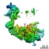
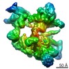
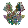
| Title | Prespliceosome structure provides insight into spliceosome assembly and regulation (map A2) | |||||||||
 Components Components |
| |||||||||
 Keywords Keywords | SPLICING / Spliceosome / A complex / RNA processing / RNA-protein complex | |||||||||
| Function / homology |  Function and homology information Function and homology informationmRNA splice site recognition / U4/U6 snRNP / 7-methylguanosine cap hypermethylation / pICln-Sm protein complex / positive regulation of mRNA splicing, via spliceosome / small nuclear ribonucleoprotein complex / SMN-Sm protein complex / spliceosomal tri-snRNP complex / splicing factor binding / snRNP binding ...mRNA splice site recognition / U4/U6 snRNP / 7-methylguanosine cap hypermethylation / pICln-Sm protein complex / positive regulation of mRNA splicing, via spliceosome / small nuclear ribonucleoprotein complex / SMN-Sm protein complex / spliceosomal tri-snRNP complex / splicing factor binding / snRNP binding / commitment complex / mRNA cis splicing, via spliceosome / U2-type prespliceosome assembly / U2-type spliceosomal complex / U1 snRNP / U2 snRNP / U4 snRNP / pre-mRNA 5'-splice site binding / U2-type prespliceosome / poly(U) RNA binding / precatalytic spliceosome / mRNA 5'-splice site recognition / spliceosomal complex assembly / Prp19 complex / U5 snRNP / spliceosomal snRNP assembly / U2 snRNA binding / U1 snRNA binding / U4/U6 x U5 tri-snRNP complex / catalytic step 2 spliceosome / spliceosomal complex / mRNA splicing, via spliceosome / response to xenobiotic stimulus / mRNA binding / RNA binding / zinc ion binding / nucleus / cytoplasm / cytosol Similarity search - Function | |||||||||
| Biological species |  | |||||||||
| Method | ELECTRON MICROSCOPY / single particle reconstruction / cryo EM / Resolution: 4 Å | |||||||||
 Authors Authors | Plaschka, C. / Lin, P.-C. / Charenton, C. / Nagai, K. | |||||||||
| Funding support |  United Kingdom, 2items United Kingdom, 2items
| |||||||||
 Citation Citation | Journal: Nature / Year: 2018 Title: Prespliceosome structure provides insights into spliceosome assembly and regulation. Authors: Clemens Plaschka / Pei-Chun Lin / Clément Charenton / Kiyoshi Nagai /  Abstract: The spliceosome catalyses the excision of introns from pre-mRNA in two steps, branching and exon ligation, and is assembled from five small nuclear ribonucleoprotein particles (snRNPs; U1, U2, U4, ...The spliceosome catalyses the excision of introns from pre-mRNA in two steps, branching and exon ligation, and is assembled from five small nuclear ribonucleoprotein particles (snRNPs; U1, U2, U4, U5, U6) and numerous non-snRNP factors. For branching, the intron 5' splice site and the branch point sequence are selected and brought by the U1 and U2 snRNPs into the prespliceosome, which is a focal point for regulation by alternative splicing factors. The U4/U6.U5 tri-snRNP subsequently joins the prespliceosome to form the complete pre-catalytic spliceosome. Recent studies have revealed the structural basis of the branching and exon-ligation reactions, however, the structural basis of the early events in spliceosome assembly remains poorly understood. Here we report the cryo-electron microscopy structure of the yeast Saccharomyces cerevisiae prespliceosome at near-atomic resolution. The structure reveals an induced stabilization of the 5' splice site in the U1 snRNP, and provides structural insights into the functions of the human alternative splicing factors LUC7-like (yeast Luc7) and TIA-1 (yeast Nam8), both of which have been linked to human disease. In the prespliceosome, the U1 snRNP associates with the U2 snRNP through a stable contact with the U2 3' domain and a transient yeast-specific contact with the U2 SF3b-containing 5' region, leaving its tri-snRNP-binding interface fully exposed. The results suggest mechanisms for 5' splice site transfer to the U6 ACAGAGA region within the assembled spliceosome and for its subsequent conversion to the activation-competent B-complex spliceosome. Taken together, the data provide a working model to investigate the early steps of spliceosome assembly. | |||||||||
| History |
|
- Structure visualization
| Movie |
Movie viewer |
|---|---|
| Structure viewer | Molecule: MolmilJmol/JSmol |
- Downloads & links
-Download
| PDBx/mmCIF format | 6g90.cif.gz | 1.6 MB | Display | PDBx/mmCIF format |
|---|---|---|---|---|
| PDB format | pdb6g90.ent.gz | 1.2 MB | Display | PDB format |
| PDBx/mmJSON format | 6g90.json.gz | Tree view | PDBx/mmJSON format | |
| Others |  Other downloads Other downloads |
-Validation report
| Arichive directory | https://data.pdbj.org/pub/pdb/validation_reports/g9/6g90ftp://data.pdbj.org/pub/pdb/validation_reports/g9/6g90 | HTTPS FTP |
|---|
-Related structure data


-Links
 PDBj
PDBj


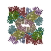
- Assembly
| Deposited unit | 
|
|---|---|
| 1 |
|
-Components
-RNA chain , 3 types, 3 molecules 12I
| #1: RNA chain | Mass: 126116.875 Da / Num. of mol.: 1 / Source method: isolated from a natural source / Source: (natural) |
|---|---|
| #2: RNA chain | Mass: 45762.836 Da / Num. of mol.: 1 / Source method: isolated from a natural source / Source: (natural) |
| #11: RNA chain | Mass: 12045.173 Da / Num. of mol.: 1 / Source method: obtained synthetically / Source: (synth.) |
-U1 small nuclear ribonucleoprotein ... , 5 types, 5 molecules ABCEJ
| #3: Protein | Mass: 33754.168 Da / Num. of mol.: 1 / Source method: isolated from a natural source / Source: (natural) |
|---|---|
| #4: Protein | Mass: 33445.242 Da / Num. of mol.: 1 / Source method: isolated from a natural source / Source: (natural) |
| #5: Protein | Mass: 27106.016 Da / Num. of mol.: 1 / Source method: isolated from a natural source / Source: (natural) |
| #7: Protein | Mass: 65222.020 Da / Num. of mol.: 1 / Source method: isolated from a natural source / Source: (natural) |
| #12: Protein | Mass: 71484.555 Da / Num. of mol.: 1 / Source method: isolated from a natural source / Source: (natural) |
-Protein , 10 types, 11 molecules DFGHOQRXZbs
| #6: Protein | Mass: 74834.742 Da / Num. of mol.: 1 / Source method: isolated from a natural source / Source: (natural) |
|---|---|
| #8: Protein | Mass: 57010.840 Da / Num. of mol.: 1 / Source method: isolated from a natural source / Source: (natural) |
| #9: Protein | Mass: 56575.277 Da / Num. of mol.: 1 / Source method: isolated from a natural source / Source: (natural) |
| #10: Protein | Mass: 28504.295 Da / Num. of mol.: 1 / Source method: isolated from a natural source / Source: (natural) |
| #13: Protein | Mass: 110166.672 Da / Num. of mol.: 1 / Source method: isolated from a natural source / Source: (natural) |
| #15: Protein | Mass: 50210.699 Da / Num. of mol.: 1 / Source method: isolated from a natural source / Source: (natural) |
| #16: Protein | Mass: 24534.152 Da / Num. of mol.: 1 / Source method: isolated from a natural source / Source: (natural) |
| #22: Protein | Mass: 4358.364 Da / Num. of mol.: 1 / Source method: isolated from a natural source / Source: (natural) |
| #24: Protein | Mass: 10045.401 Da / Num. of mol.: 1 / Source method: isolated from a natural source / Source: (natural) |
| #25: Protein | Mass: 22426.990 Da / Num. of mol.: 2 / Source method: isolated from a natural source / Source: (natural) |
-Pre-mRNA-splicing factor ... , 5 types, 5 molecules PSTUV
| #14: Protein | Mass: 153956.781 Da / Num. of mol.: 1 / Source method: isolated from a natural source / Source: (natural) |
|---|---|
| #17: Protein | Mass: 12283.573 Da / Num. of mol.: 1 / Source method: isolated from a natural source / Source: (natural) |
| #18: Protein | Mass: 63126.445 Da / Num. of mol.: 1 / Source method: isolated from a natural source / Source: (natural) |
| #19: Protein | Mass: 29292.230 Da / Num. of mol.: 1 / Source method: isolated from a natural source / Source: (natural) |
| #20: Protein | Mass: 33111.512 Da / Num. of mol.: 1 / Source method: isolated from a natural source / Source: (natural) |
-U2 small nuclear ribonucleoprotein ... , 2 types, 2 molecules WY
| #21: Protein | Mass: 27232.252 Da / Num. of mol.: 1 / Source method: isolated from a natural source / Source: (natural) |
|---|---|
| #23: Protein | Mass: 12850.944 Da / Num. of mol.: 1 / Source method: isolated from a natural source / Source: (natural) |
-Small nuclear ribonucleoprotein ... , 6 types, 12 molecules dvewfxgyhtiu
| #26: Protein | Mass: 11240.139 Da / Num. of mol.: 2 / Source method: isolated from a natural source / Source: (natural) #27: Protein | Mass: 10385.098 Da / Num. of mol.: 2 / Source method: isolated from a natural source / Source: (natural) #28: Protein | Mass: 9669.945 Da / Num. of mol.: 2 / Source method: isolated from a natural source / Source: (natural) #29: Protein | Mass: 8490.809 Da / Num. of mol.: 2 / Source method: isolated from a natural source / Source: (natural) #30: Protein | Mass: 16296.798 Da / Num. of mol.: 2 / Source method: isolated from a natural source / Source: (natural) #31: Protein | Mass: 12876.066 Da / Num. of mol.: 2 / Source method: isolated from a natural source / Source: (natural) |
|---|
-Non-polymers , 1 types, 9 molecules 
| #32: Chemical | ChemComp-ZN /  Mass: 65.409 Da / Num. of mol.: 9 / Source method: obtained synthetically / Formula: Zn Mass: 65.409 Da / Num. of mol.: 9 / Source method: obtained synthetically / Formula: Zn |
|---|
-Details
| Has protein modification | Y |
|---|
-Experimental details
-Experiment
| Experiment | Method: ELECTRON MICROSCOPY |
|---|---|
| EM experiment | Aggregation state: PARTICLE / 3D reconstruction method: single particle reconstruction |
- Sample preparation
| Component | Name: Prespliceosome A complex / Type: COMPLEX / Entity ID: #1-#31 / Source: NATURAL | |||||||||||||||||||||||||||||||||||
|---|---|---|---|---|---|---|---|---|---|---|---|---|---|---|---|---|---|---|---|---|---|---|---|---|---|---|---|---|---|---|---|---|---|---|---|---|
| Molecular weight | Value: 1.8 MDa / Experimental value: YES | |||||||||||||||||||||||||||||||||||
| Source (natural) | Organism: | |||||||||||||||||||||||||||||||||||
| Buffer solution | pH: 7.9 | |||||||||||||||||||||||||||||||||||
| Buffer component |
| |||||||||||||||||||||||||||||||||||
| Specimen | Conc.: 0.4 mg/ml / Embedding applied: NO / Shadowing applied: NO / Staining applied: NO / Vitrification applied: YES | |||||||||||||||||||||||||||||||||||
| Specimen support | Grid material: COPPER / Grid mesh size: 400 divisions/in. / Grid type: Quantifoil R2/2 | |||||||||||||||||||||||||||||||||||
| Vitrification | Instrument: FEI VITROBOT MARK III / Cryogen name: ETHANE / Humidity: 100 % Details: Grids were blotted for 2-3.5 s and vitrified by plunging into liquid ethane with a FEI Vitrobot Mark III operated at 4 degree Celsius and 100% humidity. |
- Electron microscopy imaging
| Experimental equipment |  Model: Titan Krios / Image courtesy: FEI Company |
|---|---|
| Microscopy | Model: FEI TITAN KRIOS |
| Electron gun | Electron source:  FIELD EMISSION GUN / Accelerating voltage: 300 kV / Illumination mode: FLOOD BEAM FIELD EMISSION GUN / Accelerating voltage: 300 kV / Illumination mode: FLOOD BEAM |
| Electron lens | Mode: BRIGHT FIELD / Nominal magnification: 105000 X / Cs: 2.7 mm |
| Specimen holder | Cryogen: NITROGEN / Specimen holder model: FEI TITAN KRIOS AUTOGRID HOLDER |
| Image recording | Electron dose: 43 e/Å2 / Detector mode: COUNTING / Film or detector model: GATAN K2 SUMMIT (4k x 4k) / Num. of real images: 9407 |
| Image scans | Movie frames/image: 20 |
- Processing
| EM software |
| ||||||||||||||||||||||||||||
|---|---|---|---|---|---|---|---|---|---|---|---|---|---|---|---|---|---|---|---|---|---|---|---|---|---|---|---|---|---|
| CTF correction | Type: PHASE FLIPPING AND AMPLITUDE CORRECTION | ||||||||||||||||||||||||||||
| Symmetry | Point symmetry: C1 (asymmetric) | ||||||||||||||||||||||||||||
| 3D reconstruction | Resolution: 4 Å / Resolution method: FSC 0.143 CUT-OFF / Num. of particles: 153556 / Symmetry type: POINT | ||||||||||||||||||||||||||||
| Atomic model building | Space: REAL |
