 Movie
Movie Controller

[English] 日本語
 Yorodumi
- PDB-6cpv: MicroED structure of NaK ion channel reveals a process of Na+ par... -
+ Open data

- Basic information
| Entry | Database: PDB / ID: 6cpv | ||||||
|---|---|---|---|---|---|---|---|
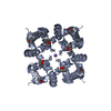
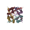
| Title | MicroED structure of NaK ion channel reveals a process of Na+ partition into the selectivity filter | ||||||
 Components Components | Potassium channel protein | ||||||
 Keywords Keywords | TRANSPORT PROTEIN / ion channel / NaK | ||||||
| Function / homology |  Function and homology information Function and homology informationpotassium channel activity / membrane / metal ion binding / identical protein binding Similarity search - Function | ||||||
| Biological species |  | ||||||
| Method | ELECTRON CRYSTALLOGRAPHY / electron crystallography /  MOLECULAR REPLACEMENT / cryo EM / Resolution: 2.5002 Å MOLECULAR REPLACEMENT / cryo EM / Resolution: 2.5002 Å | ||||||
 Authors Authors | Liu, S. / Gonen, T. | ||||||
| Funding support |  United States, 1items United States, 1items
| ||||||
 Citation Citation | Journal: Commun Biol / Year: 2018 Title: MicroED structure of the NaK ion channel reveals a Na partition process into the selectivity filter. Authors: Shian Liu / Tamir Gonen / Abstract: Sodium (Na) is a ubiquitous and important inorganic salt mediating many critical biological processes such as neuronal excitation, signaling, and facilitation of various transporters. The hydration ...Sodium (Na) is a ubiquitous and important inorganic salt mediating many critical biological processes such as neuronal excitation, signaling, and facilitation of various transporters. The hydration states of Na are proposed to play critical roles in determining the conductance and the selectivity of Na channels, yet they are rarely captured by conventional structural biology means. Here we use the emerging cryo-electron microscopy (cryoEM) method micro-electron diffraction (MicroED) to study the structure of a prototypical tetrameric Na-conducting channel, NaK, to 2.5 Å resolution from nano-crystals. Two new conformations at the external site of NaK are identified, allowing us to visualize a partially hydrated Na ion at the entrance of the channel pore. A process of dilation coupled with Na movement is identified leading to valuable insights into the mechanism of ion conduction and gating. This study lays the ground work for future studies using MicroED in membrane protein biophysics. | ||||||
| History |
|
- Structure visualization
| Movie |
Movie viewer |
|---|---|
| Structure viewer | Molecule: MolmilJmol/JSmol |
- Downloads & links
-Download
| PDBx/mmCIF format | 6cpv.cif.gz | 52.2 KB | Display | PDBx/mmCIF format |
|---|---|---|---|---|
| PDB format | pdb6cpv.ent.gz | 35.2 KB | Display | PDB format |
| PDBx/mmJSON format | 6cpv.json.gz | Tree view | PDBx/mmJSON format | |
| Others |  Other downloads Other downloads |
-Validation report
| Arichive directory | https://data.pdbj.org/pub/pdb/validation_reports/cp/6cpvftp://data.pdbj.org/pub/pdb/validation_reports/cp/6cpv | HTTPS FTP |
|---|
-Related structure data


-Links
 PDBj
PDBj

- Assembly
| Deposited unit | 
| |||||||||||||||||||||||||||||||||||||||
|---|---|---|---|---|---|---|---|---|---|---|---|---|---|---|---|---|---|---|---|---|---|---|---|---|---|---|---|---|---|---|---|---|---|---|---|---|---|---|---|---|
| 1 | 
| |||||||||||||||||||||||||||||||||||||||
| 2 | 
| |||||||||||||||||||||||||||||||||||||||
| Unit cell |
| |||||||||||||||||||||||||||||||||||||||
| Components on special symmetry positions |
|
-Components
| #1: Protein | Mass: 10706.538 Da / Num. of mol.: 2 / Fragment: UNP residues 19-110 Source method: isolated from a genetically manipulated source Source: (gene. exp.) Gene: A9485_19160, B4155_3291, BACERE00184_02078, CN419_22740, CN950_06075, CN980_22870, COI98_17615, COK18_26145, CON37_12595 Production host: #2: Chemical | ChemComp-NA /   Mass: 22.990 Da / Num. of mol.: 12 / Source method: obtained synthetically / Formula: Na Mass: 22.990 Da / Num. of mol.: 12 / Source method: obtained synthetically / Formula: Na#3: Chemical | ChemComp-MPD / ( |   Mass: 118.174 Da / Num. of mol.: 1 / Source method: obtained synthetically / Formula: C6H14O2 / Comment: precipitant*YM Mass: 118.174 Da / Num. of mol.: 1 / Source method: obtained synthetically / Formula: C6H14O2 / Comment: precipitant*YM#4: Water | ChemComp-HOH / |  Mass: 18.015 Da / Num. of mol.: 4 / Source method: isolated from a natural source / Formula: H2O Mass: 18.015 Da / Num. of mol.: 4 / Source method: isolated from a natural source / Formula: H2O |
|---|
-Experimental details
-Experiment
| Experiment | Method: ELECTRON CRYSTALLOGRAPHY |
|---|---|
| EM experiment | Aggregation state: 3D ARRAY / 3D reconstruction method: electron crystallography |
- Sample preparation
| Component | Name: NaK / Type: COMPLEX / Entity ID: #1-#2 / Source: RECOMBINANT |
|---|---|
| Source (natural) | Organism: |
| Source (recombinant) | Organism: |
| Buffer solution | pH: 7 |
| Specimen | Embedding applied: NO / Shadowing applied: NO / Staining applied: NO / Vitrification applied: YES |
| Vitrification | Cryogen name: ETHANE |
-Data collection
| Experimental equipment |  Model: Tecnai F20 / Image courtesy: FEI Company |
|---|---|
| Microscopy | Model: FEI TECNAI F20 |
| Electron gun | Electron source: FIELD EMISSION GUN / Accelerating voltage: 200 kV / Illumination mode: FLOOD BEAM |
| Electron lens | Mode: DIFFRACTION |
| Image recording | Electron dose: 0.1 e/Å2 / Film or detector model: TVIPS TEMCAM-F416 (4k x 4k) |
| EM diffraction | Camera length: 1750 mm |
| EM diffraction shell | Resolution: 2.5002→3.1486 Å / Fourier space coverage: 0.76 % / Multiplicity: 4.1 / Num. of structure factors: 2685 / Phase residual: 22.68 ° |
| EM diffraction stats | Fourier space coverage: 81.7 % / High resolution: 2.5002 Å / Num. of intensities measured: 27479 / Num. of structure factors: 5643 / Phase error: 20.27 ° / Phase residual: 20.27 ° / Phase error rejection criteria: 0 / Rmerge: 0.206 / Rsym: 0.206 |
- Processing
| Software |
| ||||||||||||||||||||||||
|---|---|---|---|---|---|---|---|---|---|---|---|---|---|---|---|---|---|---|---|---|---|---|---|---|---|
| EM 3D crystal entity | ∠α: 90 ° / ∠β: 90 ° / ∠γ: 90 ° / A: 68.0716 Å / B: 68.0716 Å / C: 89.3 Å / Space group name: I4 / Space group num: 79 | ||||||||||||||||||||||||
| CTF correction | Type: NONE | ||||||||||||||||||||||||
| 3D reconstruction | Resolution: 2.5002 Å / Resolution method: DIFFRACTION PATTERN/LAYERLINES / Symmetry type: 3D CRYSTAL | ||||||||||||||||||||||||
| Atomic model building | B value: 41 | ||||||||||||||||||||||||
| Refinement | Method to determine structure: MOLECULAR REPLACEMENT Starting model: PDB entry 3E89 Resolution: 2.5→21.992 Å / SU ML: 0.37 / Cross valid method: THROUGHOUT / σ(F): 1.41 / Phase error: 20.26
| ||||||||||||||||||||||||
| Solvent computation | Shrinkage radii: 0.9 Å / VDW probe radii: 1.11 Å | ||||||||||||||||||||||||
| Refine LS restraints |
| ||||||||||||||||||||||||
| LS refinement shell |
|
