 ムービー
ムービー コントローラー


+ データを開く

- 基本情報
基本情報
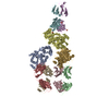
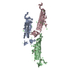
| 登録情報 | データベース: PDB / ID: 5h53 | ||||||
|---|---|---|---|---|---|---|---|
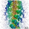
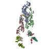
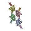
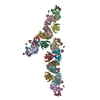
| タイトル | The structure of rabbit skeletal muscle actomyosin rigor complex at 5.2 angstrom. | ||||||
 要素 要素 |
| ||||||
 キーワード キーワード | MOTOR PROTEIN / Actin / Myosin / Muscle / rigor complex | ||||||
| 機能・相同性 |  機能・相同性情報 機能・相同性情報myosin complex / structural constituent of muscle / cytoskeletal motor activator activity / myofibril / tropomyosin binding / mesenchyme migration / troponin I binding / myosin heavy chain binding / cytoskeletal motor activity / filamentous actin ...myosin complex / structural constituent of muscle / cytoskeletal motor activator activity / myofibril / tropomyosin binding / mesenchyme migration / troponin I binding / myosin heavy chain binding / cytoskeletal motor activity / filamentous actin / actin filament bundle / skeletal muscle thin filament assembly / striated muscle thin filament / actin filament bundle assembly / skeletal muscle myofibril / actin monomer binding / skeletal muscle fiber development / stress fiber / titin binding / cellular response to starvation / actin filament polymerization / filopodium / actin filament / 加水分解酵素; 酸無水物に作用; 酸無水物に作用・細胞または細胞小器官の運動に関与 / calcium-dependent protein binding / actin filament binding / lamellipodium / cell body / hydrolase activity / protein domain specific binding / calcium ion binding / positive regulation of gene expression / magnesium ion binding / ATP binding / identical protein binding / cytoplasm 類似検索 - 分子機能 | ||||||
| 生物種 |  | ||||||
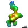
| 手法 | 電子顕微鏡法 / らせん対称体再構成法 / クライオ電子顕微鏡法 / 解像度: 5.2 Å | ||||||
 データ登録者 データ登録者 | Fujii, T. / Namba, K. | ||||||
 引用 引用 | ジャーナル: Nat Commun / 年: 2017 タイトル: Structure of actomyosin rigour complex at 5.2 Å resolution and insights into the ATPase cycle mechanism. 著者: Takashi Fujii / Keiichi Namba /  要旨: Muscle contraction is driven by cyclic association and dissociation of myosin head of the thick filament with thin actin filament coupled with ATP binding and hydrolysis by myosin. However, because ...Muscle contraction is driven by cyclic association and dissociation of myosin head of the thick filament with thin actin filament coupled with ATP binding and hydrolysis by myosin. However, because of the absence of actomyosin rigour structure at high resolution, it still remains unclear how the strong binding of myosin to actin filament triggers the release of hydrolysis products and how ATP binding causes their dissociation. Here we report the structure of mammalian skeletal muscle actomyosin rigour complex at 5.2 Å resolution by electron cryomicroscopy. Comparison with the structures of myosin in various states shows a distinctly large conformational change, providing insights into the ATPase-coupled reaction cycle of actomyosin. Based on our observations, we hypothesize that asymmetric binding along the actin filament could function as a Brownian ratchet by favouring directionally biased thermal motions of myosin and actin. | ||||||
| 履歴 |
|
- 構造の表示
構造の表示
| ムービー |
ムービービューア |
|---|---|
| 構造ビューア | 分子: MolmilJmol/JSmol |
- ダウンロードとリンク
-ダウンロード
| PDBx/mmCIF形式 | 5h53.cif.gz | 328.6 KB | 表示 | PDBx/mmCIF形式 |
|---|---|---|---|---|
| PDB形式 | pdb5h53.ent.gz | 265.6 KB | 表示 | PDB形式 |
| PDBx/mmJSON形式 | 5h53.json.gz | ツリー表示 | PDBx/mmJSON形式 | |
| その他 |  その他のダウンロード その他のダウンロード |
-検証レポート
| 文書・要旨 | 5h53_validation.pdf.gz | 1.1 MB | 表示 | wwPDB検証レポート |
|---|---|---|---|---|
| 文書・詳細版 | 5h53_full_validation.pdf.gz | 1.3 MB | 表示 | |
| XML形式データ | 5h53_validation.xml.gz | 93.3 KB | 表示 | |
| CIF形式データ | 5h53_validation.cif.gz | 132.6 KB | 表示 | |
| アーカイブディレクトリ | https://data.pdbj.org/pub/pdb/validation_reports/h5/5h53ftp://data.pdbj.org/pub/pdb/validation_reports/h5/5h53 | HTTPS FTP |
-関連構造データ
-リンク
 PDBj
PDBj


- 集合体
集合体
| 登録構造単位 | 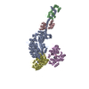
|
|---|---|
| 1 |
|
-要素
| #1: タンパク質 | 分子量: 96770.133 Da / 分子数: 1 / 断片: UNP residues 1-845 / 由来タイプ: 天然 / 由来: (天然) | ||
|---|---|---|---|
| #2: タンパク質 | 分子量: 16507.588 Da / 分子数: 1 / 断片: UNP residues 25-170 / 由来タイプ: 天然 / 由来: (天然) | ||
| #3: タンパク質 | 分子量: 17187.293 Da / 分子数: 1 / 断片: UNP residues 41-192 / 由来タイプ: 天然 / 由来: (天然) | ||
| #4: タンパク質 | 分子量: 41875.633 Da / 分子数: 2 / 断片: UNP residues 3-377 / 由来タイプ: 天然 / 由来: (天然) #5: 化合物 |   分子量: 427.201 Da / 分子数: 2 / 由来タイプ: 合成 / 式: C10H15N5O10P2 / コメント: ADP, エネルギー貯蔵分子*YM 分子量: 427.201 Da / 分子数: 2 / 由来タイプ: 合成 / 式: C10H15N5O10P2 / コメント: ADP, エネルギー貯蔵分子*YM |
-実験情報
-実験
| 実験 | 手法: 電子顕微鏡法 |
|---|---|
| EM実験 | 試料の集合状態: FILAMENT / 3次元再構成法: らせん対称体再構成法 |
- 試料調製
試料調製
| 構成要素 | 名称: Actomyosin rigor complex / タイプ: COMPLEX / Entity ID: #1-#4 / 由来: NATURAL |
|---|---|
| 由来(天然) | 生物種: |
| 緩衝液 | pH: 7.5 |
| 試料 | 包埋: NO / シャドウイング: NO / 染色: NO / 凍結: YES |
| 急速凍結 | 凍結剤: ETHANE |
- 電子顕微鏡撮影
| 顕微鏡 | モデル: JEOL 3200FSC |
|---|---|
| 電子銃 | 電子線源:  FIELD EMISSION GUN / 加速電圧: 200 kV / 照射モード: FLOOD BEAM FIELD EMISSION GUN / 加速電圧: 200 kV / 照射モード: FLOOD BEAM |
| 電子レンズ | モード: BRIGHT FIELD |
| 撮影 | 電子線照射量: 20 e/Å2 フィルム・検出器のモデル: TVIPS TEMCAM-F415 (4k x 4k) |
- 解析
解析
| CTF補正 | タイプ: PHASE FLIPPING AND AMPLITUDE CORRECTION |
|---|---|
| らせん対称 | 回転角度/サブユニット: 166.7 ° / 軸方向距離/サブユニット: 27.6 Å / らせん対称軸の対称性: C1 |
| 3次元再構成 | 解像度: 5.2 Å / 解像度の算出法: FSC 0.143 CUT-OFF / 粒子像の数: 31535 / 対称性のタイプ: HELICAL |
