 Movie
Movie Controller

[English] 日本語
 Yorodumi
- PDB-5ft2: Sub-tomogram averaging of Lassa virus glycoprotein spike from vir... -
+ Open data

- Basic information
| Entry | Database: PDB / ID: 5ft2 | |||||||||
|---|---|---|---|---|---|---|---|---|---|---|
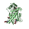
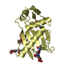
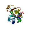
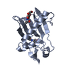
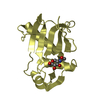
| Title | Sub-tomogram averaging of Lassa virus glycoprotein spike from virus- like particles at pH 5 | |||||||||
 Components Components | PRE-GLYCOPROTEIN POLYPROTEIN GP COMPLEX | |||||||||
 Keywords Keywords | CELL ADHESION / MEMBRANE PROTEIN / GLYCOPROTEIN / RECEPTOR BINDING / MEMBRANE FUSION | |||||||||
| Function / homology |  Function and homology information Function and homology informationhost cell Golgi membrane / receptor-mediated endocytosis of virus by host cell / host cell endoplasmic reticulum membrane / fusion of virus membrane with host endosome membrane / viral envelope / virion attachment to host cell / host cell plasma membrane / virion membrane / metal ion binding / membrane Similarity search - Function | |||||||||
| Biological species |  LASSA VIRUS LASSA VIRUS | |||||||||
| Method | ELECTRON MICROSCOPY / electron tomography / cryo EM / Resolution: 16.4 Å | |||||||||
 Authors Authors | Li, S. / Zhaoyang, S. / Pryce, R. / Parsy, M.L. / Fehling, S.K. / Schlie, K. / Siebert, C.A. / Garten, W. / Bowden, T.A. / Strecker, T. / Huiskonen, J.T. | |||||||||
 Citation Citation | Journal: PLoS Pathog / Year: 2016 Title: Acidic pH-Induced Conformations and LAMP1 Binding of the Lassa Virus Glycoprotein Spike. Authors: Sai Li / Zhaoyang Sun / Rhys Pryce / Marie-Laure Parsy / Sarah K Fehling / Katrin Schlie / C Alistair Siebert / Wolfgang Garten / Thomas A Bowden / Thomas Strecker / Juha T Huiskonen /   Abstract: Lassa virus is an enveloped, bi-segmented RNA virus and the most prevalent and fatal of all Old World arenaviruses. Virus entry into the host cell is mediated by a tripartite surface spike complex, ...Lassa virus is an enveloped, bi-segmented RNA virus and the most prevalent and fatal of all Old World arenaviruses. Virus entry into the host cell is mediated by a tripartite surface spike complex, which is composed of two viral glycoprotein subunits, GP1 and GP2, and the stable signal peptide. Of these, GP1 binds to cellular receptors and GP2 catalyzes fusion between the viral envelope and the host cell membrane during endocytosis. The molecular structure of the spike and conformational rearrangements induced by low pH, prior to fusion, remain poorly understood. Here, we analyzed the three-dimensional ultrastructure of Lassa virus using electron cryotomography. Sub-tomogram averaging yielded a structure of the glycoprotein spike at 14-Å resolution. The spikes are trimeric, cover the virion envelope, and connect to the underlying matrix. Structural changes to the spike, following acidification, support a viral entry mechanism dependent on binding to the lysosome-resident receptor LAMP1 and further dissociation of the membrane-distal GP1 subunits. #1: Journal: J Virol / Year: 2015Title: Molecular Mechanism for LAMP1 Recognition by Lassa Virus. Authors: Hadas Cohen-Dvashi / Nadav Cohen / Hadar Israeli / Ron Diskin /  Abstract: Lassa virus is a notorious human pathogen that infects many thousands of people each year in West Africa, causing severe viral hemorrhagic fevers and significant mortality. The surface glycoprotein ...Lassa virus is a notorious human pathogen that infects many thousands of people each year in West Africa, causing severe viral hemorrhagic fevers and significant mortality. The surface glycoprotein of Lassa virus mediates receptor recognition through its GP1 subunit. Here we report the crystal structure of GP1 from Lassa virus, which is the first representative GP1 structure for Old World arenaviruses. We identify a unique triad of histidines that forms a binding site for LAMP1, a known lysosomal protein recently discovered to be a critical receptor for internalized Lassa virus at acidic pH. We demonstrate that mutation of this histidine triad, which is highly conserved among Old World arenaviruses, impairs LAMP1 recognition. Our biochemical and structural data further suggest that GP1 from Lassa virus may undergo irreversible conformational changes that could serve as an immunological decoy mechanism. Together with a variable region that we identify on the surface of GP1, those could be two distinct mechanisms that Lassa virus utilizes to avoid antibody-based immune response. IMPORTANCE: Structural data at atomic resolution for viral proteins is key for understanding their function at the molecular level and can facilitate novel avenues for combating viral infections. ...IMPORTANCE: Structural data at atomic resolution for viral proteins is key for understanding their function at the molecular level and can facilitate novel avenues for combating viral infections. Here we used X-ray protein crystallography to decipher the crystal structure of the receptor-binding domain (GP1) from Lassa virus. This is a pathogenic virus that causes significant illness and mortality in West Africa. This structure reveals the overall architecture of GP1 domains from the group of viruses known as the Old World arenaviruses. Using this structural information, we elucidated the mechanisms for pH switch and binding of Lassa virus to LAMP1, a recently identified host receptor that is critical for successful infection. Lastly, our structural analysis suggests two novel immune evasion mechanisms that Lassa virus may utilize to escape antibody-based immune response. | |||||||||
| History |
|
- Structure visualization
| Movie |
Movie viewer |
|---|---|
| Structure viewer | Molecule: MolmilJmol/JSmol |
- Downloads & links
-Download
| PDBx/mmCIF format | 5ft2.cif.gz | 79.5 KB | Display | PDBx/mmCIF format |
|---|---|---|---|---|
| PDB format | pdb5ft2.ent.gz | 58.2 KB | Display | PDB format |
| PDBx/mmJSON format | 5ft2.json.gz | Tree view | PDBx/mmJSON format | |
| Others |  Other downloads Other downloads |
-Validation report
| Arichive directory | https://data.pdbj.org/pub/pdb/validation_reports/ft/5ft2ftp://data.pdbj.org/pub/pdb/validation_reports/ft/5ft2 | HTTPS FTP |
|---|
-Related structure data
| Related structure data | 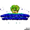 3292MC 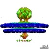 3290C  3291C 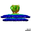 3293C  3294C M: map data used to model this data C: citing same article ( |
|---|---|
| Similar structure data |


-Links
 PDBj
PDBj- Assembly
| Deposited unit | 
|
|---|---|
| 1 |
|
-Components
| #1: Protein | Mass: 19379.693 Da / Num. of mol.: 1 / Fragment: RECEPTOR BINDING DOMAIN, UNP RESIDUES 75-237 Source method: isolated from a genetically manipulated source Source: (gene. exp.) LASSA VIRUS / Strain: JOSIAH / Production host:  TRICHOPLUSIA NI (cabbage looper) / References: UniProt: P08669 TRICHOPLUSIA NI (cabbage looper) / References: UniProt: P08669 | ||||||
|---|---|---|---|---|---|---|---|
| #2: Polysaccharide | Source method: isolated from a genetically manipulated source #3: Sugar |   Type: D-saccharide, beta linking / Mass: 221.208 Da / Num. of mol.: 2 Type: D-saccharide, beta linking / Mass: 221.208 Da / Num. of mol.: 2Source method: isolated from a genetically manipulated source Formula: C8H15NO6 #4: Water | ChemComp-HOH / |  Mass: 18.015 Da / Num. of mol.: 23 / Source method: isolated from a natural source / Formula: H2O Mass: 18.015 Da / Num. of mol.: 23 / Source method: isolated from a natural source / Formula: H2OHas protein modification | Y | |
-Experimental details
-Experiment
| Experiment | Method: ELECTRON MICROSCOPY |
|---|---|
| EM experiment | Aggregation state: PARTICLE / 3D reconstruction method: electron tomography |
- Sample preparation
| Component | Name: PURIFIED LASSA VIRUS VLPS AT PH 5 / Type: VIRUS |
|---|---|
| Buffer solution | Name: 50 MM BUFFER OF SUCCINIC ACID, DIHYDROGEN PHOSPHATE AND GLYCINE (SPG) pH: 5.2 Details: 50 MM BUFFER OF SUCCINIC ACID, DIHYDROGEN PHOSPHATE AND GLYCINE (SPG) |
| Specimen | Embedding applied: NO / Shadowing applied: NO / Staining applied: NO / Vitrification applied: YES |
| Specimen support | Details: HOLEY CARBON |
| Vitrification | Instrument: GATAN CRYOPLUNGE 3 / Cryogen name: ETHANE-PROPANE Details: VITRIFICATION 1 -- CRYOGEN- ETHANE-PROPANE MIXTURE, HUMIDITY- 80, TEMPERATURE- 120, INSTRUMENT- GATAN CRYOPLUNGE 3, METHOD- BLOT FOR 3 SECONDS BEFORE PLUNGING., |
- Electron microscopy imaging
| Experimental equipment |  Model: Tecnai F30 / Image courtesy: FEI Company |
|---|---|
| Microscopy | Model: FEI TECNAI F30 / Date: Sep 25, 2014 / Details: SUPER-RESOLUTION COUNTING MODE |
| Electron gun | Electron source:  FIELD EMISSION GUN / Accelerating voltage: 300 kV / Illumination mode: FLOOD BEAM FIELD EMISSION GUN / Accelerating voltage: 300 kV / Illumination mode: FLOOD BEAM |
| Electron lens | Mode: BRIGHT FIELD / Nominal magnification: 160000 X / Calibrated magnification: 37037 X / Nominal defocus max: 6700 nm / Nominal defocus min: 2800 nm / Cs: 2 mm |
| Specimen holder | Tilt angle max: 45 ° / Tilt angle min: -45 ° |
| Image recording | Electron dose: 60 e/Å2 / Film or detector model: GATAN K2 SUMMIT (4k x 4k) |
| Image scans | Num. digital images: 16 |
- Processing
| EM software |
| ||||||||||||||||||||
|---|---|---|---|---|---|---|---|---|---|---|---|---|---|---|---|---|---|---|---|---|---|
| CTF correction | Details: EACH TILTED IMAGE | ||||||||||||||||||||
| Symmetry | Point symmetry: C3 (3 fold cyclic) | ||||||||||||||||||||
| 3D reconstruction | Method: TEMPLATE BASED / Resolution: 16.4 Å / Num. of particles: 2578 / Nominal pixel size: 2.7 Å / Actual pixel size: 2.7 Å Details: SUB-TOMOGRAM AVERAGING SUBMISSION BASED ON EXPERIMENTAL DATA FROM EMDB EMD-3292. (DEPOSITION ID: 14158). Symmetry type: POINT | ||||||||||||||||||||
| Atomic model building | Protocol: RIGID BODY FIT / Space: REAL / Target criteria: Cross-correlation coefficient / Details: METHOD--RIGID BODY REFINEMENT PROTOCOL--X-RAY | ||||||||||||||||||||
| Atomic model building | PDB-ID: 4ZJF Accession code: 4ZJF / Source name: PDB / Type: experimental model | ||||||||||||||||||||
| Refinement | Highest resolution: 16.4 Å | ||||||||||||||||||||
| Refinement step | Cycle: LAST / Highest resolution: 16.4 Å
|

