Movie
Movie Controller

[English] 日本語
 Yorodumi
- PDB-5ahv: Cryo-EM structure of helical ANTH and ENTH tubules on PI(4,5)P2-c... -
+ Open data

- Basic information
| Entry | Database: PDB / ID: 5ahv | |||||||||
|---|---|---|---|---|---|---|---|---|---|---|
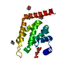
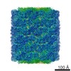
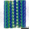
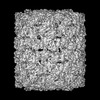
| Title | Cryo-EM structure of helical ANTH and ENTH tubules on PI(4,5)P2-containing membranes | |||||||||
 Components Components |
| |||||||||
 Keywords Keywords | CLATHRIN-BINDING PROTEIN / CLATHRIN BINDING PROTEIN / EPSIN / HIP1R / ENTH / CLATHRIN ADAPTORS / ENDOCYTOSIS | |||||||||
| Function / homology |  Function and homology information Function and homology informationCargo recognition for clathrin-mediated endocytosis / actin cortical patch assembly / clathrin vesicle coat / clathrin light chain binding / incipient cellular bud site / actin cortical patch / cellular bud tip / negative regulation of Arp2/3 complex-mediated actin nucleation / clathrin coat assembly / clathrin-cargo adaptor activity ...Cargo recognition for clathrin-mediated endocytosis / actin cortical patch assembly / clathrin vesicle coat / clathrin light chain binding / incipient cellular bud site / actin cortical patch / cellular bud tip / negative regulation of Arp2/3 complex-mediated actin nucleation / clathrin coat assembly / clathrin-cargo adaptor activity / cellular bud neck / mating projection tip / phosphatidylinositol-3,4-bisphosphate binding / phosphatidylinositol-3,5-bisphosphate binding / clathrin-coated vesicle / K63-linked polyubiquitin modification-dependent protein binding / cortical actin cytoskeleton / clathrin binding / actin filament organization / ubiquitin binding / phospholipid binding / endocytosis / actin filament binding / early endosome / endosome / plasma membrane / cytoplasm Similarity search - Function | |||||||||
| Biological species |  | |||||||||
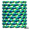
| Method | ELECTRON MICROSCOPY / single particle reconstruction / cryo EM / Resolution: 13.6 Å | |||||||||
 Authors Authors | Skruzny, M. / Desfosses, A. / Prinz, S. / Dodonova, S.O. / Gieras, A. / Uetrecht, C. / Jakobi, A.J. / Abella, M. / Hagen, W.J.H. / Schulz, J. ...Skruzny, M. / Desfosses, A. / Prinz, S. / Dodonova, S.O. / Gieras, A. / Uetrecht, C. / Jakobi, A.J. / Abella, M. / Hagen, W.J.H. / Schulz, J. / Meijers, R. / Rybin, V. / Briggs, J.A.G. / Sachse, C. / Kaksonen, M. | |||||||||
 Citation Citation | Journal: Dev Cell / Year: 2015 Title: An organized co-assembly of clathrin adaptors is essential for endocytosis. Authors: Michal Skruzny / Ambroise Desfosses / Simone Prinz / Svetlana O Dodonova / Anna Gieras / Charlotte Uetrecht / Arjen J Jakobi / Marc Abella / Wim J H Hagen / Joachim Schulz / Rob Meijers / ...Authors: Michal Skruzny / Ambroise Desfosses / Simone Prinz / Svetlana O Dodonova / Anna Gieras / Charlotte Uetrecht / Arjen J Jakobi / Marc Abella / Wim J H Hagen / Joachim Schulz / Rob Meijers / Vladimir Rybin / John A G Briggs / Carsten Sachse / Marko Kaksonen /  Abstract: Clathrin-mediated endocytosis, the main trafficking route from the plasma membrane to the cytoplasm, is critical to many fundamental cellular processes. Clathrin, coupled to the membrane by adaptor ...Clathrin-mediated endocytosis, the main trafficking route from the plasma membrane to the cytoplasm, is critical to many fundamental cellular processes. Clathrin, coupled to the membrane by adaptor proteins, is thought to play a major structural role in endocytosis by self-assembling into a cage-like lattice around the forming vesicle. Although clathrin adaptors are essential for endocytosis, little is known about their structural role in this process. Here we show that the membrane-binding domains of two conserved clathrin adaptors, Sla2 and Ent1, co-assemble in a PI(4,5)P2-dependent manner to form organized lattices on membranes. We determined the structure of the co-assembled lattice by electron cryo-microscopy and designed mutations that specifically impair the lattice formation in vitro. We show that these mutations block endocytosis in vivo. We suggest that clathrin adaptors not only link the polymerized clathrin to the membrane but also form an oligomeric structure, which is essential for membrane remodeling during endocytosis. | |||||||||
| History |
|
- Structure visualization
| Movie |
Movie viewer |
|---|---|
| Structure viewer | Molecule: MolmilJmol/JSmol |
- Downloads & links
-Download
| PDBx/mmCIF format | 5ahv.cif.gz | 148.7 KB | Display | PDBx/mmCIF format |
|---|---|---|---|---|
| PDB format | pdb5ahv.ent.gz | 116.7 KB | Display | PDB format |
| PDBx/mmJSON format | 5ahv.json.gz | Tree view | PDBx/mmJSON format | |
| Others |  Other downloads Other downloads |
-Validation report
| Arichive directory | https://data.pdbj.org/pub/pdb/validation_reports/ah/5ahvftp://data.pdbj.org/pub/pdb/validation_reports/ah/5ahv | HTTPS FTP |
|---|
-Related structure data
| Related structure data | 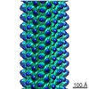 2896MC 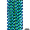 2897C M: map data used to model this data C: citing same article ( |
|---|---|
| Similar structure data |

-Links
 PDBj
PDBj


- Assembly
| Deposited unit | 
|
|---|---|
| 1 | x 78
|
-Components
| #1: Protein | Mass: 18005.471 Da / Num. of mol.: 1 / Fragment: RESIDUES 5-272 Source method: isolated from a genetically manipulated source Source: (gene. exp.) Plasmid: PETM30 / Production host:  |
|---|---|
| #2: Protein | Mass: 30700.961 Da / Num. of mol.: 1 / Fragment: RESIDUES 1-154 Source method: isolated from a genetically manipulated source Source: (gene. exp.) Plasmid: PETM30 / Production host: |
-Experimental details
-Experiment
| Experiment | Method: ELECTRON MICROSCOPY |
|---|---|
| EM experiment | Aggregation state: FILAMENT / 3D reconstruction method: single particle reconstruction |
- Sample preparation
| Component | Name: ANTH AND ENTH DOMAINS OF THE CLATHRIN ADAPTORS SLA2 AND ENT1, RESPECTIVELY Type: ORGANELLE OR CELLULAR COMPONENT |
|---|---|
| Buffer solution | Name: 20 MM HEPES, PH 7.5, 100 MM KCL / pH: 7.5 / Details: 20 MM HEPES, PH 7.5, 100 MM KCL |
| Specimen | Embedding applied: NO / Shadowing applied: NO / Staining applied: NO / Vitrification applied: YES |
| Specimen support | Details: HOLEY CARBON |
| Vitrification | Instrument: HOMEMADE PLUNGER / Cryogen name: ETHANE Details: VITRIFICATION 1 -- CRYOGEN- ETHANE, HUMIDITY- 80, INSTRUMENT- HOMEMADE PLUNGER, METHOD- , APPLIED ON C-FLAT HOLEY CARBON COATED GRIDS (PROTOCHIPS) AND VITRIFIED, |
- Electron microscopy imaging
| Experimental equipment |  Model: Titan Krios / Image courtesy: FEI Company |
|---|---|
| Microscopy | Model: FEI TITAN KRIOS / Date: Mar 1, 2013 |
| Electron gun | Electron source:  FIELD EMISSION GUN / Accelerating voltage: 120 kV / Illumination mode: FLOOD BEAM FIELD EMISSION GUN / Accelerating voltage: 120 kV / Illumination mode: FLOOD BEAM |
| Electron lens | Mode: BRIGHT FIELD / Nominal magnification: 59000 X / Calibrated magnification: 59000 X / Nominal defocus max: 5000 nm / Nominal defocus min: 500 nm / Cs: 2.7 mm |
| Image recording | Electron dose: 10 e/Å2 / Film or detector model: GATAN ULTRASCAN 4000 (4k x 4k) |
| Image scans | Num. digital images: 1419 |
- Processing
| EM software |
| |||||||||||||||
|---|---|---|---|---|---|---|---|---|---|---|---|---|---|---|---|---|
| CTF correction | Details: EACH PARTICLE | |||||||||||||||
| 3D reconstruction | Resolution: 13.6 Å / Num. of particles: 457344 / Nominal pixel size: 1.78 Å / Actual pixel size: 1.78 Å Details: SUBMISSION BASED ON EXPERIMENTAL DATA FROM EMDB EMD-2896. (DEPOSITION ID: 13053). Symmetry type: HELICAL | |||||||||||||||
| Atomic model building | Protocol: FLEXIBLE FIT / Space: REAL Details: METHOD--FLEXIBLE REFINEMENT PROTOCOL--HOMOLOGY MODEL | |||||||||||||||
| Atomic model building | PDB-ID: 1H0A Accession code: 1H0A / Source name: PDB / Type: experimental model | |||||||||||||||
| Refinement | Highest resolution: 13.6 Å | |||||||||||||||
| Refinement step | Cycle: LAST / Highest resolution: 13.6 Å
|