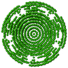
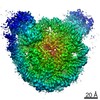
ジャーナル: J Mol Biol / 年: 2013 タイトル: Location of the dsRNA-dependent polymerase, VP1, in rotavirus particles. 著者: Leandro F Estrozi / Ethan C Settembre / Gaël Goret / Brian McClain / Xing Zhang / James Z Chen / Nikolaus Grigorieff / Stephen C Harrison / 要旨: Double-stranded RNA (dsRNA) viruses transcribe and replicate RNA within an assembled, inner capsid particle; only plus-sense mRNA emerges into the intracellular milieu. During infectious entry of a ...Double-stranded RNA (dsRNA) viruses transcribe and replicate RNA within an assembled, inner capsid particle; only plus-sense mRNA emerges into the intracellular milieu. During infectious entry of a rotavirus particle, the outer layer of its three-layer structure dissociates, delivering the inner double-layered particle (DLP) into the cytosol. DLP structures determined by X-ray crystallography and electron cryomicroscopy (cryoEM) show that the RNA coils uniformly into the particle interior, avoiding a "fivefold hub" of more structured density projecting inward from the VP2 shell of the DLP along each of the twelve 5-fold axes. Analysis of the X-ray crystallographic electron density map suggested that principal contributors to the hub are the N-terminal arms of VP2, but reexamination of the cryoEM map has shown that many features come from a molecule of VP1, randomly occupying five equivalent and partly overlapping positions. We confirm here that the electron density in the X-ray map leads to the same conclusion, and we describe the functional implications of the orientation and position of the polymerase. The exit channel for the nascent transcript directs the nascent transcript toward an opening along the 5-fold axis. The template strand enters from within the particle, and the dsRNA product of the initial replication step exits in a direction tangential to the inner surface of the VP2 shell, allowing it to coil optimally within the DLP. The polymerases of reoviruses appear to have similar positions and functional orientations.
モード: BRIGHT FIELD / 倍率(公称値): 59000 X / 倍率(補正後): 56540 X / 最大 デフォーカス(公称値): 3500 nm / 最小 デフォーカス(公称値): 1100 nm / Cs: 2 mm
試料ホルダ
温度: 90 K / 傾斜角・最大: 0.001 ° / 傾斜角・最小: 0 °
撮影
電子線照射量: 15 e/Å2 / フィルム・検出器のモデル: KODAK SO-163 FILM
画像スキャン
デジタル画像の数: 386
放射波長
相対比: 1
-
解析
EMソフトウェア
ID
名称
カテゴリ
1
URO
モデルフィッティング
2
VEDA
モデルフィッティング
3
FPM
3次元再構成
4
RICO
3次元再構成
CTF補正
詳細: INDIVIDUAL PARTICLE PHASE FLIPPING
対称性
点対称性: I (正20面体型対称)
3次元再構成
手法: SYMMETRY-ADAPTED FUNCTIONS / 解像度: 6 Å / 粒子像の数: 7000 / ピクセルサイズ(公称値): 1.18 Å / ピクセルサイズ(実測値): 1.23 Å 詳細: SUBMISSION BASED ON EXPERIMENTAL DATA FROM EMDB EMD-2100.(DEPOSITION ID: 10810). 対称性のタイプ: POINT
 ムービー
ムービー コントローラー


 データを開く
 基本情報
基本情報 要素
要素 キーワード
キーワード 機能・相同性情報
 BOVINE ROTAVIRUS (ウイルス)
 データ登録者
 引用
引用
 構造の表示
構造の表示 ダウンロードとリンク
 その他のダウンロード


 PDBj
PDBj 集合体
集合体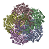
 試料調製
試料調製 電子顕微鏡撮影
 FIELD EMISSION GUN / 加速電圧: 300 kV / 照射モード: FLOOD BEAM
 解析
解析
