 ムービー
ムービー コントローラー


+ データを開く

- 基本情報
基本情報
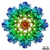
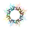
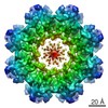
| 登録情報 | データベース: PDB / ID: 3jd6 | ||||||
|---|---|---|---|---|---|---|---|
| タイトル | Double octamer structure of retinoschisin, a cell-cell adhesion protein of the retina | ||||||
 要素 要素 | Retinoschisin | ||||||
 キーワード キーワード | CELL ADHESION / discoidin domain / double octamer / adhesion protein / X-linked retinoschisis | ||||||
| 機能・相同性 |  機能・相同性情報 機能・相同性情報neuron to neuron synapse / retina layer formation / eye development / phosphatidylinositol-3,4-bisphosphate binding / phosphatidylserine binding / photoreceptor inner segment / visual perception / protein homooligomerization / cell adhesion / external side of plasma membrane ...neuron to neuron synapse / retina layer formation / eye development / phosphatidylinositol-3,4-bisphosphate binding / phosphatidylserine binding / photoreceptor inner segment / visual perception / protein homooligomerization / cell adhesion / external side of plasma membrane / protein-containing complex binding / protein-containing complex / extracellular space 類似検索 - 分子機能 | ||||||
| 生物種 |  Homo sapiens (ヒト) Homo sapiens (ヒト) | ||||||
| 手法 | 電子顕微鏡法 / 単粒子再構成法 / クライオ電子顕微鏡法 / 解像度: 4.1 Å | ||||||
 データ登録者 データ登録者 | Tolun, G. / Vijayasarathy, C. / Huang, R. / Zeng, Y. / Li, Y. / Steven, A.C. / Sieving, P.A. / Heymann, J.B. | ||||||
 引用 引用 | ジャーナル: Proc Natl Acad Sci U S A / 年: 2016 タイトル: Paired octamer rings of retinoschisin suggest a junctional model for cell-cell adhesion in the retina. 著者: Gökhan Tolun / Camasamudram Vijayasarathy / Rick Huang / Yong Zeng / Yan Li / Alasdair C Steven / Paul A Sieving / J Bernard Heymann /  要旨: Retinoschisin (RS1) is involved in cell-cell junctions in the retina, but is unique among known cell-adhesion proteins in that it is a soluble secreted protein. Loss-of-function mutations in RS1 lead ...Retinoschisin (RS1) is involved in cell-cell junctions in the retina, but is unique among known cell-adhesion proteins in that it is a soluble secreted protein. Loss-of-function mutations in RS1 lead to early vision impairment in young males, called X-linked retinoschisis. The disease is characterized by separation of inner retinal layers and disruption of synaptic signaling. Using cryo-electron microscopy, we report the structure at 4.1 Å, revealing double octamer rings not observed before. Each subunit is composed of a discoidin domain and a small N-terminal (RS1) domain. The RS1 domains occupy the centers of the rings, but are not required for ring formation and are less clearly defined, suggesting mobility. We determined the structure of the discoidin rings, consistent with known intramolecular and intermolecular disulfides. The interfaces internal to and between rings feature residues implicated in X-linked retinoschisis, indicating the importance of correct assembly. Based on this structure, we propose that RS1 couples neighboring membranes together through octamer-octamer contacts, perhaps modulated by interactions with other membrane components. | ||||||
| 履歴 |
|
- 構造の表示
構造の表示
| ムービー |
ムービービューア |
|---|---|
| 構造ビューア | 分子: MolmilJmol/JSmol |
- ダウンロードとリンク
-ダウンロード
| PDBx/mmCIF形式 | 3jd6.cif.gz | 68.5 KB | 表示 | PDBx/mmCIF形式 |
|---|---|---|---|---|
| PDB形式 | pdb3jd6.ent.gz | 49.7 KB | 表示 | PDB形式 |
| PDBx/mmJSON形式 | 3jd6.json.gz | ツリー表示 | PDBx/mmJSON形式 | |
| その他 |  その他のダウンロード その他のダウンロード |
-検証レポート
| 文書・要旨 | 3jd6_validation.pdf.gz | 925.5 KB | 表示 | wwPDB検証レポート |
|---|---|---|---|---|
| 文書・詳細版 | 3jd6_full_validation.pdf.gz | 925.6 KB | 表示 | |
| XML形式データ | 3jd6_validation.xml.gz | 16.3 KB | 表示 | |
| CIF形式データ | 3jd6_validation.cif.gz | 22.2 KB | 表示 | |
| アーカイブディレクトリ | https://data.pdbj.org/pub/pdb/validation_reports/jd/3jd6ftp://data.pdbj.org/pub/pdb/validation_reports/jd/3jd6 | HTTPS FTP |
-関連構造データ

-リンク
 PDBj
PDBj

- 集合体
集合体
| 登録構造単位 | 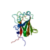
|
|---|---|
| 1 | x 16
|
| 2 |
|
| 3 | 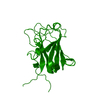
|
| 対称性 | 点対称性: (シェーンフリース記号: D8 (2回x8回 2面回転対称)) |
-要素
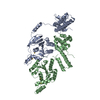
| #1: タンパク質 | 分子量: 23889.830 Da / 分子数: 1 / 断片: UNP residues 24-224 / 由来タイプ: 組換発現 / 由来: (組換発現) Homo sapiens (ヒト) / 遺伝子: RS1, XLRS1 / プラスミド: 11109-E01発現宿主:   Spodoptera frugiperda (ツマジロクサヨトウ) Spodoptera frugiperda (ツマジロクサヨトウ)株 (発現宿主): Sf9 / 参照: UniProt: O15537 |
|---|---|
| Has protein modification | Y |
-実験情報
-実験
| 実験 | 手法: 電子顕微鏡法 |
|---|---|
| EM実験 | 試料の集合状態: PARTICLE / 3次元再構成法: 単粒子再構成法 |
- 試料調製
試料調製
| 構成要素 | 名称: Purified Retinoschisin: residues 24-224 + 6xHis / タイプ: COMPLEX / 別称: RS1 |
|---|---|
| 緩衝液 | 名称: 20 mM Tris-HCl, 150 mM NaCl / pH: 7.5 / 詳細: 20 mM Tris-HCl, 150 mM NaCl |
| 試料 | 濃度: 0.2 mg/ml / 包埋: NO / シャドウイング: NO / 染色: NO / 凍結: YES |
| 試料支持 | 詳細: Plasma-cleaned 2.0 um hole, 2.0 um space C-flat holey carbon grids (Protochips) |
| 急速凍結 | 装置: LEICA EM GP / 凍結剤: ETHANE / 湿度: 90 % 詳細: Ambient temperature 23 degrees C. Plunged into liquid ethane (LEICA EM GP). |
- 電子顕微鏡撮影
| 実験機器 |  モデル: Tecnai Polara / 画像提供: FEI Company |
|---|---|
| 顕微鏡 | モデル: FEI POLARA 300 / 日付: 2014年6月3日 / 詳細: 10 frames over 3 seconds |
| 電子銃 | 電子線源:  FIELD EMISSION GUN / 加速電圧: 300 kV / 照射モード: FLOOD BEAM FIELD EMISSION GUN / 加速電圧: 300 kV / 照射モード: FLOOD BEAM |
| 電子レンズ | モード: BRIGHT FIELD / 倍率(公称値): 39000 X / Cs: 2 mm |
| 試料ホルダ | 試料ホルダーモデル: OTHER / 資料ホルダタイプ: FEI Polara specimen loader |
| 撮影 | 電子線照射量: 30 e/Å2 / フィルム・検出器のモデル: GATAN K2 (4k x 4k) / 詳細: Counting mode |
| 画像スキャン | デジタル画像の数: 19 |
- 解析
解析
| EMソフトウェア |
| ||||||||||||||||||||||||
|---|---|---|---|---|---|---|---|---|---|---|---|---|---|---|---|---|---|---|---|---|---|---|---|---|---|
| CTF補正 | 詳細: Included in iterative refinement | ||||||||||||||||||||||||
| 対称性 | 点対称性: D8 (2回x8回 2面回転対称) | ||||||||||||||||||||||||
| 3次元再構成 | 手法: Bayesian orientation refinement with iterative gridded reconstruction 解像度: 4.1 Å / 解像度の算出法: FSC 0.143 CUT-OFF / 粒子像の数: 9096 / ピクセルサイズ(公称値): 1.03 Å / ピクセルサイズ(実測値): 1.03 Å / 対称性のタイプ: POINT | ||||||||||||||||||||||||
| 原子モデル構築 | プロトコル: FLEXIBLE FIT / 空間: REAL / Target criteria: FSC(0.5) = 4.7 詳細: METHOD--Flexible fitting REFINEMENT PROTOCOL--FLEXIBLE | ||||||||||||||||||||||||
| 原子モデル構築 | PDB-ID: 1SDD Accession code: 1SDD / Source name: PDB / タイプ: experimental model | ||||||||||||||||||||||||
| 精密化ステップ | サイクル: LAST
|

