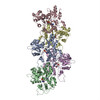
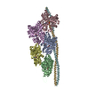
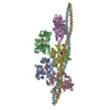
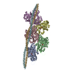
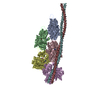
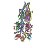
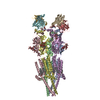
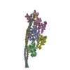
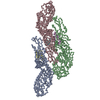
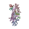
ジャーナル: Nature / 年: 2015 タイトル: Structure of the F-actin-tropomyosin complex. 著者: Julian von der Ecken / Mirco Müller / William Lehman / Dietmar J Manstein / Pawel A Penczek / Stefan Raunser / 要旨: Filamentous actin (F-actin) is the major protein of muscle thin filaments, and actin microfilaments are the main component of the eukaryotic cytoskeleton. Mutations in different actin isoforms lead ...Filamentous actin (F-actin) is the major protein of muscle thin filaments, and actin microfilaments are the main component of the eukaryotic cytoskeleton. Mutations in different actin isoforms lead to early-onset autosomal dominant non-syndromic hearing loss, familial thoracic aortic aneurysms and dissections, and multiple variations of myopathies. In striated muscle fibres, the binding of myosin motors to actin filaments is mainly regulated by tropomyosin and troponin. Tropomyosin also binds to F-actin in smooth muscle and in non-muscle cells and stabilizes and regulates the filaments there in the absence of troponin. Although crystal structures for monomeric actin (G-actin) are available, a high-resolution structure of F-actin is still missing, hampering our understanding of how disease-causing mutations affect the function of thin muscle filaments and microfilaments. Here we report the three-dimensional structure of F-actin at a resolution of 3.7 Å in complex with tropomyosin at a resolution of 6.5 Å, determined by electron cryomicroscopy. The structure reveals that the D-loop is ordered and acts as a central region for hydrophobic and electrostatic interactions that stabilize the F-actin filament. We clearly identify map density corresponding to ADP and Mg(2+) and explain the possible effect of prominent disease-causing mutants. A comparison of F-actin with G-actin reveals the conformational changes during filament formation and identifies the D-loop as their key mediator. We also confirm that negatively charged tropomyosin interacts with a positively charged groove on F-actin. Comparison of the position of tropomyosin in F-actin-tropomyosin with its position in our previously determined F-actin-tropomyosin-myosin structure reveals a myosin-induced transition of tropomyosin. Our results allow us to understand the role of individual mutations in the genesis of actin- and tropomyosin-related diseases and will serve as a strong foundation for the targeted development of drugs.
らせん対称: (回転対称性: 1 / Dyad axis: no / N subunits divisor: 1 / Num. of operations: 1 / Rise per n subunits: 27.5 Å / Rotation per n subunits: -166.4 °)
非結晶学的対称性 (NCS)
NCSドメイン:
ID
Ens-ID
詳細
1
1
chainA
2
1
chainB
3
1
chainC
4
1
chainD
5
1
chainE
NCSドメイン領域:
Dom-ID
Component-ID
Ens-ID
Selection details
Auth asym-ID
Auth seq-ID
1
1
1
chainA
A
5 - 371
2
1
1
chainB
B
5 - 371
3
1
1
chainC
C
5 - 371
4
1
1
chainD
D
5 - 371
5
1
1
chainE
E
5 - 371
詳細
The full filament can be generated using the provided helical parameters from a small repeating subunit comprising chain A and residues 145-184 of chains F and G.
-
要素
#1: タンパク質
tropomyosinalpha-1
分子量: 11507.176 Da / 分子数: 2 / 断片: SEE REMARK 999 / 由来タイプ: 組換発現 / 由来: (組換発現) Mus musculus (ハツカネズミ) / 発現宿主: Escherichia coli (大腸菌) / キーワード: SEE REMARK 999
MOUSE TROPOMYSIN WAS USED (UNP P58771, RESIDUES 97-231). DUE TO THE LIMITED RESOLUTION OF THE CRYO- ...MOUSE TROPOMYSIN WAS USED (UNP P58771, RESIDUES 97-231). DUE TO THE LIMITED RESOLUTION OF THE CRYO-EM DENSITY IN THE REGION OF TROPOMYOSIN, TROPOMYOSIN HAS BEEN REPRESENTED AS POLY(UNK).
-
実験情報
-
実験
実験
手法: 電子顕微鏡法
EM実験
試料の集合状態: FILAMENT / 3次元再構成法: らせん対称体再構成法
-
試料調製
構成要素
ID
名称
タイプ
詳細
Parent-ID
1
F-actin-tropomyosin complex
COMPLEX
Filament
0
2
F-actin (alpha)
1
3
tropomyosin (alpha)
1
緩衝液
名称: 5 mM Tris-HCl, pH 7.5, 1 mM DTT, 100 mM KCl, 2 mM MgCl2 pH: 7.5 詳細: 5 mM Tris-HCl, pH 7.5, 1 mM DTT, 100 mM KCl, 2 mM MgCl2
装置: GATAN CRYOPLUNGE 3 / 凍結剤: ETHANE / Temp: 106 K / 湿度: 90 % 詳細: Sample was applied to grid, incubated for 10 seconds, and manually blotted for 3 seconds from the backside with filter paper before plunging into liquid ethane (GATAN CRYOPLUNGE 3) 手法: Sample was applied to grid, incubated for 10 seconds, and manually blotted for 3 seconds from the backside with filter paper.
手法: helicon, cter / 解像度: 3.7 Å / 解像度の算出法: FSC 0.5 CUT-OFF / ピクセルサイズ(公称値): 1.14 Å / ピクセルサイズ(実測値): 1.12 Å 詳細: The tropomyosin map filtered to 6.5 Angstrom was merged with the final F-actin map (3.7 Angstrom) to obtain a map of the entire F-actin tropomyosin complex. Coordinates must be shifted by (0. ...詳細: The tropomyosin map filtered to 6.5 Angstrom was merged with the final F-actin map (3.7 Angstrom) to obtain a map of the entire F-actin tropomyosin complex. Coordinates must be shifted by (0.123 -0.381 0.273) to match coordinates to fibre diffraction standards (filament axis = z axis passing through x=0 and y=0). 対称性のタイプ: HELICAL
 ムービー
ムービー コントローラー


 データを開く
 基本情報
基本情報 要素
要素 キーワード
キーワード 機能・相同性情報
 データ登録者
 引用
引用

 構造の表示
構造の表示 ダウンロードとリンク
 その他のダウンロード
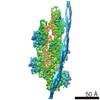
 PDBj
PDBj


 集合体
集合体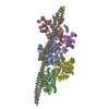


 分子量: 24.305 Da / 分子数: 5 / 由来タイプ: 合成 / 式: Mg
 分子量: 427.201 Da / 分子数: 5 / 由来タイプ: 合成 / 式: C10H15N5O10P2 / コメント: ADP, エネルギー貯蔵分子*YM
 試料調製
試料調製 電子顕微鏡撮影
 FIELD EMISSION GUN / 加速電圧: 300 kV / 照射モード: OTHER
 解析
解析