 ムービー
ムービー コントローラー


+ データを開く

- 基本情報
基本情報
| 登録情報 | データベース: PDB / ID: 3j6e | ||||||
|---|---|---|---|---|---|---|---|
| タイトル | Energy minimized average structure of Microtubules stabilized by GmpCpp | ||||||
 要素 要素 |
| ||||||
 キーワード キーワード | STRUCTURAL PROTEIN / microtubule / GmpCpp | ||||||
| 機能・相同性 |  機能・相同性情報 機能・相同性情報加水分解酵素; 酸無水物に作用; GTPに作用・細胞または細胞小器官の運動に関与 / structural constituent of cytoskeleton / microtubule cytoskeleton organization / mitotic cell cycle / microtubule / hydrolase activity / GTPase activity / GTP binding / metal ion binding / cytoplasm 類似検索 - 分子機能 | ||||||
| 生物種 |  | ||||||
| 手法 | 電子顕微鏡法 / 単粒子再構成法 / クライオ電子顕微鏡法 / 解像度: 4.7 Å | ||||||
 データ登録者 データ登録者 | Alushin, G.M. / Lander, G.C. / Kellogg, E.H. / Zhang, R. / Baker, D. / Nogales, E. | ||||||
 引用 引用 | ジャーナル: Cell / 年: 2014 タイトル: High-resolution microtubule structures reveal the structural transitions in αβ-tubulin upon GTP hydrolysis. 著者: Gregory M Alushin / Gabriel C Lander / Elizabeth H Kellogg / Rui Zhang / David Baker / Eva Nogales /  要旨: Dynamic instability, the stochastic switching between growth and shrinkage, is essential for microtubule function. This behavior is driven by GTP hydrolysis in the microtubule lattice and is ...Dynamic instability, the stochastic switching between growth and shrinkage, is essential for microtubule function. This behavior is driven by GTP hydrolysis in the microtubule lattice and is inhibited by anticancer agents like Taxol. We provide insight into the mechanism of dynamic instability, based on high-resolution cryo-EM structures (4.7-5.6 Å) of dynamic microtubules and microtubules stabilized by GMPCPP or Taxol. We infer that hydrolysis leads to a compaction around the E-site nucleotide at longitudinal interfaces, as well as movement of the α-tubulin intermediate domain and H7 helix. Displacement of the C-terminal helices in both α- and β-tubulin subunits suggests an effect on interactions with binding partners that contact this region. Taxol inhibits most of these conformational changes, allosterically inducing a GMPCPP-like state. Lateral interactions are similar in all conditions we examined, suggesting that microtubule lattice stability is primarily modulated at longitudinal interfaces. | ||||||
| 履歴 |
|
- 構造の表示
構造の表示
| ムービー |
ムービービューア |
|---|---|
| 構造ビューア | 分子: MolmilJmol/JSmol |
- ダウンロードとリンク
-ダウンロード
| PDBx/mmCIF形式 | 3j6e.cif.gz | 1.3 MB | 表示 | PDBx/mmCIF形式 |
|---|---|---|---|---|
| PDB形式 | pdb3j6e.ent.gz | 1 MB | 表示 | PDB形式 |
| PDBx/mmJSON形式 | 3j6e.json.gz | ツリー表示 | PDBx/mmJSON形式 | |
| その他 |  その他のダウンロード その他のダウンロード |
-検証レポート
| 文書・要旨 | 3j6e_validation.pdf.gz | 2.1 MB | 表示 | wwPDB検証レポート |
|---|---|---|---|---|
| 文書・詳細版 | 3j6e_full_validation.pdf.gz | 2.2 MB | 表示 | |
| XML形式データ | 3j6e_validation.xml.gz | 211.5 KB | 表示 | |
| CIF形式データ | 3j6e_validation.cif.gz | 304.7 KB | 表示 | |
| アーカイブディレクトリ | https://data.pdbj.org/pub/pdb/validation_reports/j6/3j6eftp://data.pdbj.org/pub/pdb/validation_reports/j6/3j6e | HTTPS FTP |
-関連構造データ


-リンク
 PDBj
PDBj


- 集合体
集合体
| 登録構造単位 | 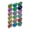
|
|---|---|
| 1 |
|
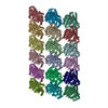
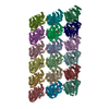
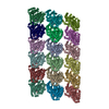
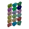
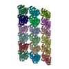
| 詳細 | The 3 x 3 lattice represented in this entry is a segment of a microtubule. |
-要素
-タンパク質 , 2種, 18分子 ACEGIKMOQBDFHJLNPR
| #1: タンパク質 | 分子量: 48769.988 Da / 分子数: 9 / 由来タイプ: 天然 / 由来: (天然) #2: タンパク質 | 分子量: 47940.945 Da / 分子数: 9 / 由来タイプ: 天然 / 由来: (天然) |
|---|
-非ポリマー , 4種, 108分子 


| #3: 化合物 | ChemComp-GTP /  分子量: 523.180 Da / 分子数: 9 / 由来タイプ: 合成 / 式: C10H16N5O14P3 / コメント: GTP, エネルギー貯蔵分子*YM 分子量: 523.180 Da / 分子数: 9 / 由来タイプ: 合成 / 式: C10H16N5O14P3 / コメント: GTP, エネルギー貯蔵分子*YM#4: 化合物 | ChemComp-MG /  分子量: 24.305 Da / 分子数: 18 / 由来タイプ: 合成 / 式: Mg 分子量: 24.305 Da / 分子数: 18 / 由来タイプ: 合成 / 式: Mg#5: 化合物 | ChemComp-G2P /  分子量: 521.208 Da / 分子数: 9 / 由来タイプ: 合成 / 式: C11H18N5O13P3 分子量: 521.208 Da / 分子数: 9 / 由来タイプ: 合成 / 式: C11H18N5O13P3コメント: GMP-CPP, エネルギー貯蔵分子類似体*YM #6: 水 | ChemComp-HOH / | 分子量: 18.015 Da / 分子数: 72 / 由来タイプ: 天然 / 式: H2O |
|---|
-実験情報
-実験
| 実験 | 手法: 電子顕微鏡法 |
|---|---|
| EM実験 | 試料の集合状態: FILAMENT / 3次元再構成法: 単粒子再構成法 |
- 試料調製
試料調製
| 構成要素 | 名称: microtubule stabilized by GMPCPP / タイプ: COMPLEX |
|---|---|
| 緩衝液 | 名称: 80 mM PIPES, 1 mM EGTA, 1 mM MgCl2, 1 mM DTT, 0.05% Nonidet P-40 pH: 6.8 詳細: 80 mM PIPES, 1 mM EGTA, 1 mM MgCl2, 1 mM DTT, 0.05% Nonidet P-40 |
| 試料 | 濃度: 0.25 mg/ml / 包埋: NO / シャドウイング: NO / 染色: NO / 凍結: YES |
| 試料支持 | 詳細: 400 mesh C-flat 1.2/1.3, glow discharged in Edwards carbon evaporator |
| 急速凍結 | 装置: FEI VITROBOT MARK II / 凍結剤: ETHANE / Temp: 90.4 K / 湿度: 90 % 詳細: The grid was blotted for 2 seconds before plunging into liquid ethane (FEI VITROBOT MARK II). 手法: The grid was blotted for 2 seconds before plunging. |
- 電子顕微鏡撮影
| 実験機器 |  モデル: Titan Krios / 画像提供: FEI Company |
|---|---|
| 顕微鏡 | モデル: FEI TITAN KRIOS / 日付: 2012年4月26日 |
| 電子銃 | 電子線源:  FIELD EMISSION GUN / 加速電圧: 300 kV / 照射モード: FLOOD BEAM FIELD EMISSION GUN / 加速電圧: 300 kV / 照射モード: FLOOD BEAM |
| 電子レンズ | モード: BRIGHT FIELD / 倍率(公称値): 72000 X / 倍率(補正後): 72000 X / 最大 デフォーカス(公称値): 3500 nm / 最小 デフォーカス(公称値): 1400 nm / Cs: 2.7 mm |
| 試料ホルダ | 試料ホルダーモデル: GATAN LIQUID NITROGEN / 資料ホルダタイプ: Gatan 626 holder / 傾斜角・最大: 0 ° / 傾斜角・最小: 0 ° |
| 撮影 | 電子線照射量: 25 e/Å2 / フィルム・検出器のモデル: KODAK SO-163 FILM |
| 画像スキャン | デジタル画像の数: 252 |
| 放射 | プロトコル: SINGLE WAVELENGTH / 単色(M)・ラウエ(L): M / 散乱光タイプ: x-ray |
| 放射波長 | 相対比: 1 |
- 解析
解析
| EMソフトウェア |
| ||||||||||||||||||||
|---|---|---|---|---|---|---|---|---|---|---|---|---|---|---|---|---|---|---|---|---|---|
| CTF補正 | 詳細: ctftilt | ||||||||||||||||||||
| 3次元再構成 | 手法: projection matching / 解像度: 4.7 Å / 解像度の算出法: FSC 0.143 CUT-OFF / 粒子像の数: 57451 / ピクセルサイズ(公称値): 1.74 Å / ピクセルサイズ(実測値): 1.74 Å / 対称性のタイプ: HELICAL | ||||||||||||||||||||
| 原子モデル構築 | プロトコル: FLEXIBLE FIT / 空間: REAL 詳細: REFINEMENT PROTOCOL--flexible fitting DETAILS--Structure represents the minimized average structure of the 1% lowest energy structures from the refinement run. | ||||||||||||||||||||
| 原子モデル構築 | PDB-ID: 1JFF Accession code: 1JFF / Source name: PDB / タイプ: experimental model | ||||||||||||||||||||
| 精密化ステップ | サイクル: LAST
|

