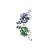
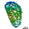
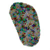
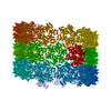
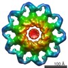
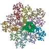
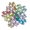
ジャーナル: Nature / 年: 2013 タイトル: Mature HIV-1 capsid structure by cryo-electron microscopy and all-atom molecular dynamics. 著者: Gongpu Zhao / Juan R Perilla / Ernest L Yufenyuy / Xin Meng / Bo Chen / Jiying Ning / Jinwoo Ahn / Angela M Gronenborn / Klaus Schulten / Christopher Aiken / Peijun Zhang / 要旨: Retroviral capsid proteins are conserved structurally but assemble into different morphologies. The mature human immunodeficiency virus-1 (HIV-1) capsid is best described by a 'fullerene cone' model, ...Retroviral capsid proteins are conserved structurally but assemble into different morphologies. The mature human immunodeficiency virus-1 (HIV-1) capsid is best described by a 'fullerene cone' model, in which hexamers of the capsid protein are linked to form a hexagonal surface lattice that is closed by incorporating 12 capsid-protein pentamers. HIV-1 capsid protein contains an amino-terminal domain (NTD) comprising seven α-helices and a β-hairpin, a carboxy-terminal domain (CTD) comprising four α-helices, and a flexible linker with a 310-helix connecting the two structural domains. Structures of the capsid-protein assembly units have been determined by X-ray crystallography; however, structural information regarding the assembled capsid and the contacts between the assembly units is incomplete. Here we report the cryo-electron microscopy structure of a tubular HIV-1 capsid-protein assembly at 8 Å resolution and the three-dimensional structure of a native HIV-1 core by cryo-electron tomography. The structure of the tubular assembly shows, at the three-fold interface, a three-helix bundle with critical hydrophobic interactions. Mutagenesis studies confirm that hydrophobic residues in the centre of the three-helix bundle are crucial for capsid assembly and stability, and for viral infectivity. The cryo-electron-microscopy structures enable modelling by large-scale molecular dynamics simulation, resulting in all-atom models for the hexamer-of-hexamer and pentamer-of-hexamer elements as well as for the entire capsid. Incorporation of pentamers results in closer trimer contacts and induces acute surface curvature. The complete atomic HIV-1 capsid model provides a platform for further studies of capsid function and for targeted pharmacological intervention.
A: capsid protein B: capsid protein C: capsid protein D: capsid protein E: capsid protein F: capsid protein G: capsid protein H: capsid protein I: capsid protein J: capsid protein K: capsid protein L: capsid protein M: capsid protein N: capsid protein O: capsid protein P: capsid protein Q: capsid protein R: capsid protein S: capsid protein T: capsid protein U: capsid protein V: capsid protein W: capsid protein X: capsid protein Y: capsid protein Z: capsid protein 5: capsid protein a: capsid protein b: capsid protein c: capsid protein 6: capsid protein i: capsid protein j: capsid protein k: capsid protein l: capsid protein m: capsid protein 7: capsid protein d: capsid protein e: capsid protein f: capsid protein g: capsid protein h: capsid protein
名称: HIV-1 capsid protein / タイプ: COMPLEX / 詳細: hexamer
分子量
値: 0.025 MDa / 実験値: NO
緩衝液
名称: 1 M NaCl, 50 mM Tris-HCl / pH: 8 / 詳細: 1 M NaCl, 50 mM Tris-HCl
試料
濃度: 2 mg/ml / 包埋: NO / シャドウイング: NO / 染色: NO / 凍結: YES
試料支持
詳細: 200 mesh quantifoil R2/1 copper grid
急速凍結
装置: HOMEMADE PLUNGER / 凍結剤: ETHANE / Temp: 110 K / 湿度: 80 % 詳細: With 2.5 uL sample on carbon side, add 3 uL dilution buffer (100 mM NaCl, 50 mM Tris, pH 8.0) to back side. Blot 3-5 seconds from back side and plunge into liquid ethane with a homemade plunger. 手法: With 2.5 uL sample on carbon side, add 3 uL dilution buffer (100 mM NaCl, 50 mM Tris, pH 8.0) to back side. Blot 3-5 seconds from back side.
モード: BRIGHT FIELD / 倍率(公称値): 59000 X / 倍率(補正後): 58257 X / 最大 デフォーカス(公称値): 3500 nm / 最小 デフォーカス(公称値): 1000 nm / Cs: 2 mm / カメラ長: 0 mm
試料ホルダ
試料ホルダーモデル: OTHER / 資料ホルダタイプ: Polara cartridge / 温度: 82 K / 最高温度: 85 K / 最低温度: 80 K / 傾斜角・最大: 0 ° / 傾斜角・最小: 0 °
撮影
電子線照射量: 15 e/Å2 / フィルム・検出器のモデル: KODAK SO-163 FILM
画像スキャン
デジタル画像の数: 27
-
解析
EMソフトウェア
ID
名称
カテゴリ
1
MDFF
モデルフィッティング
2
FREALIGN
3次元再構成
CTF補正
詳細: each filament
らせん対称
回転角度/サブユニット: 31.13 ° / 軸方向距離/サブユニット: 7.247 Å / らせん対称軸の対称性: C1
3次元再構成
手法: real space helical reconstruction / 解像度: 8.6 Å / 解像度の算出法: FSC 0.5 CUT-OFF / 粒子像の数: 3210 / ピクセルサイズ(実測値): 1.09 Å 詳細: (Helical Details: The segments were aligned and reconstructed using Frealign. Twofold symmetry was imposed using IHRSR++.) 対称性のタイプ: HELICAL
原子モデル構築
ID
プロトコル
空間
詳細
1
FLEXIBLEFIT
REAL
METHOD--The MDFF-derived HOH structure was equilibrated in 1 M NaCl FOR 425 ns using MD without applying restraints. PDB entries 3H47 AND 2KOD were the starting structures. REFINEMENT PROTOCOL--flexible
2
FLEXIBLEFIT
REAL
METHOD--The MDFF-derived HOH structure was equilibrated in 1 M NaCl FOR 425 ns using MD without applying restraints. PDB entries 3H47 AND 2KOD were the starting structures. REFINEMENT PROTOCOL--flexible
 ムービー
ムービー コントローラー


 データを開く
 基本情報
基本情報 要素
要素 キーワード
キーワード 機能・相同性情報
 Human immunodeficiency virus 1 (ヒト免疫不全ウイルス)
 データ登録者
 引用
引用
 構造の表示
構造の表示 ダウンロードとリンク
 その他のダウンロード
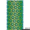


 PDBj
PDBj

 集合体
集合体

 試料調製
試料調製 電子顕微鏡撮影
 FIELD EMISSION GUN / 加速電圧: 200 kV / 照射モード: FLOOD BEAM
 解析
解析