 Movie
Movie Controller

[English] 日本語
 Yorodumi
- PDB-3j0i: Fitting of the phiKZ gp29PR structure into the cryo-EM density ma... -
+ Open data

- Basic information
| Entry | Database: PDB / ID: 3j0i | ||||||
|---|---|---|---|---|---|---|---|
| Title | Fitting of the phiKZ gp29PR structure into the cryo-EM density map of the phiKZ polysheath | ||||||
 Components Components | PHIKZ029 | ||||||
 Keywords Keywords | STRUCTURAL PROTEIN / bacteriophage tail sheath | ||||||
| Function / homology | : / Bacteriophage phiKZ, gp29PR / PHIKZ029 Function and homology information Function and homology information | ||||||
| Biological species |  Pseudomonas phage phiKZ (virus) Pseudomonas phage phiKZ (virus) | ||||||
| Method | ELECTRON MICROSCOPY / single particle reconstruction / cryo EM / Resolution: 19 Å | ||||||
 Authors Authors | Aksyuk, A.A. / Fokine, A. / Kurochkina, L.P. / Mesyanzhinov, V.V. / Rossmann, M.G. | ||||||
 Citation Citation | Journal: Structure / Year: 2011 Title: Structural conservation of the myoviridae phage tail sheath protein fold. Authors: Anastasia A Aksyuk / Lidia P Kurochkina / Andrei Fokine / Farhad Forouhar / Vadim V Mesyanzhinov / Liang Tong / Michael G Rossmann /  Abstract: Bacteriophage phiKZ is a giant phage that infects Pseudomonas aeruginosa, a human pathogen. The phiKZ virion consists of a 1450 Å diameter icosahedral head and a 2000 Å-long contractile tail. The ...Bacteriophage phiKZ is a giant phage that infects Pseudomonas aeruginosa, a human pathogen. The phiKZ virion consists of a 1450 Å diameter icosahedral head and a 2000 Å-long contractile tail. The structure of the whole virus was previously reported, showing that its tail organization in the extended state is similar to the well-studied Myovirus bacteriophage T4 tail. The crystal structure of a tail sheath protein fragment of phiKZ was determined to 2.4 Å resolution. Furthermore, crystal structures of two prophage tail sheath proteins were determined to 1.9 and 3.3 Å resolution. Despite low sequence identity between these proteins, all of these structures have a similar fold. The crystal structure of the phiKZ tail sheath protein has been fitted into cryo-electron-microscopy reconstructions of the extended tail sheath and of a polysheath. The structural rearrangement of the phiKZ tail sheath contraction was found to be similar to that of phage T4. | ||||||
| History |
|
- Structure visualization
| Movie |
Movie viewer |
|---|---|
| Structure viewer | Molecule: MolmilJmol/JSmol |
- Downloads & links
-Download
| PDBx/mmCIF format | 3j0i.cif.gz | 577.7 KB | Display | PDBx/mmCIF format |
|---|---|---|---|---|
| PDB format | pdb3j0i.ent.gz | 480.9 KB | Display | PDB format |
| PDBx/mmJSON format | 3j0i.json.gz | Tree view | PDBx/mmJSON format | |
| Others |  Other downloads Other downloads |
-Validation report
| Arichive directory | https://data.pdbj.org/pub/pdb/validation_reports/j0/3j0iftp://data.pdbj.org/pub/pdb/validation_reports/j0/3j0i | HTTPS FTP |
|---|
-Related structure data
| Related structure data | 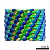 5331MC 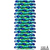 5332C 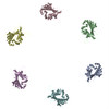 3j0hC 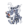 3speC C: citing same article ( M: map data used to model this data |
|---|---|
| Similar structure data |


-Links
 PDBj
PDBj- Assembly
| Deposited unit | 
|
|---|---|
| 1 |
|
-Components
| #1: Protein | Mass: 32296.910 Da / Num. of mol.: 6 Fragment: protease-resistant fragment of gene product 29 (GP29PR, UNP residues 96-390) Source method: isolated from a natural source / Source: (natural) Pseudomonas phage phiKZ (virus) / References: UniProt: Q8SDD3 |
|---|
-Experimental details
-Experiment
| Experiment | Method: ELECTRON MICROSCOPY |
|---|---|
| EM experiment | Aggregation state: PARTICLE / 3D reconstruction method: single particle reconstruction |
- Sample preparation
| Component | Name: Bacteriophage phiKZ polysheath / Type: VIRUS / Details: tail portion of the virion was selected |
|---|---|
| Details of virus | Host category: BACTERIA(EUBACTERIA) / Isolate: SPECIES / Type: VIRION |
| Natural host | Organism: Pseudomonas aeruginosa |
| Specimen | Embedding applied: NO / Shadowing applied: NO / Staining applied: NO / Vitrification applied: YES |
| Vitrification | Cryogen name: ETHANE / Details: liquid ethane |
- Electron microscopy imaging
| Microscopy | Model: FEI/PHILIPS CM200FEG |
|---|---|
| Electron gun | Electron source:  FIELD EMISSION GUN / Accelerating voltage: 200 kV / Illumination mode: SPOT SCAN FIELD EMISSION GUN / Accelerating voltage: 200 kV / Illumination mode: SPOT SCAN |
| Electron lens | Mode: BRIGHT FIELD |
| Radiation | Protocol: SINGLE WAVELENGTH / Monochromatic (M) / Laue (L): M / Scattering type: x-ray |
| Radiation wavelength | Relative weight: 1 |
- Processing
| EM software |
| ||||||||||||
|---|---|---|---|---|---|---|---|---|---|---|---|---|---|
| Symmetry | Point symmetry: C6 (6 fold cyclic) | ||||||||||||
| 3D reconstruction | Method: spider single particle / Resolution: 19 Å / Resolution method: FSC 0.5 CUT-OFF / Nominal pixel size: 2.49 Å / Actual pixel size: 2.49 Å / Details: FSC at 0.5 cut-off / Symmetry type: POINT | ||||||||||||
| Atomic model building | Space: REAL | ||||||||||||
| Atomic model building | PDB-ID: 3SPE Accession code: 3SPE / Source name: PDB / Type: experimental model | ||||||||||||
| Refinement step | Cycle: LAST
|
