 Movie
Movie Controller


+ Open data

- Basic information
| Entry | Database: EMDB / ID: EMD-5331 | |||||||||
|---|---|---|---|---|---|---|---|---|---|---|
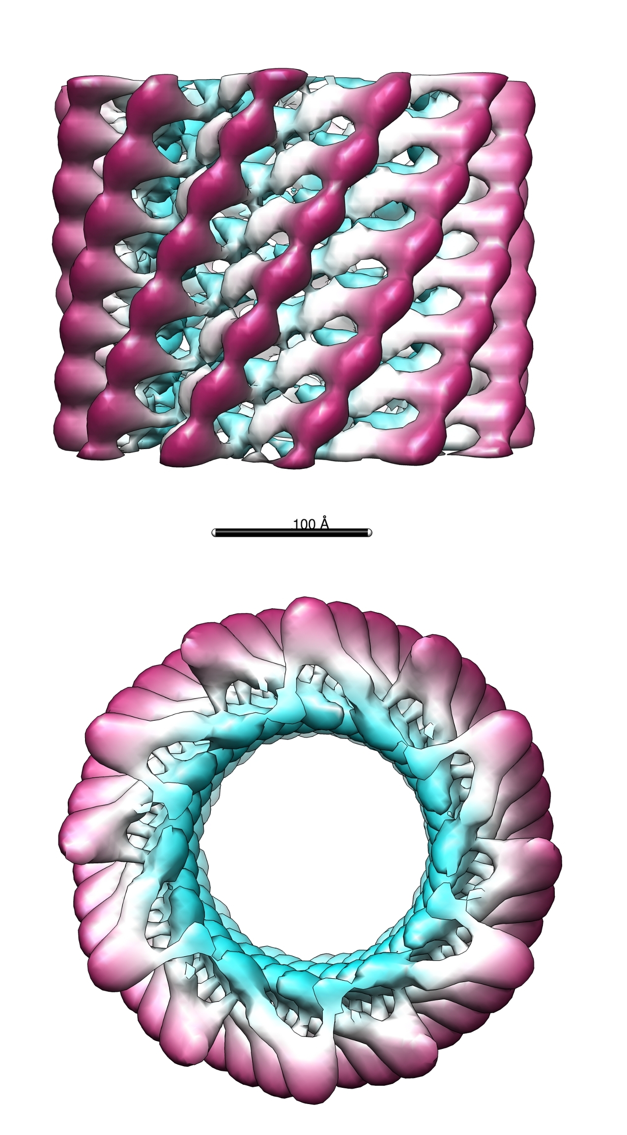
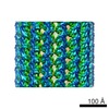
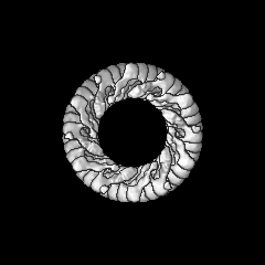
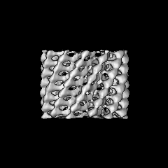
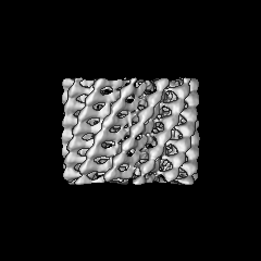
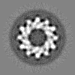
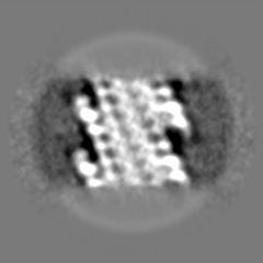
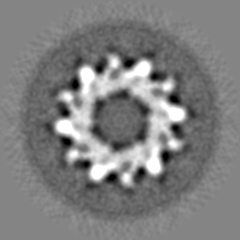
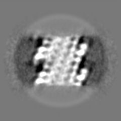
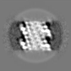
| Title | The cryoEM structure of the bacteriophage phiKZ polysheath | |||||||||
 Map data Map data | This is a map of the bacteriophage phiKZ polysheath | |||||||||
 Sample Sample |
| |||||||||
 Keywords Keywords | homopolymer of the bacteriophage tail sheath protein | |||||||||
| Function / homology | : / Bacteriophage phiKZ, gp29PR / PHIKZ029 Function and homology information Function and homology information | |||||||||
| Biological species |  Pseudomonas phage phiKZ (virus) Pseudomonas phage phiKZ (virus) | |||||||||
| Method | single particle reconstruction / cryo EM / Resolution: 19.0 Å | |||||||||
 Authors Authors | Aksyuk AA / Kurochkina LP / Fokine A / Mesyanzhinov VV / Rossmann MG | |||||||||
 Citation Citation | Journal: Structure / Year: 2011 Title: Structural conservation of the myoviridae phage tail sheath protein fold. Authors: Anastasia A Aksyuk / Lidia P Kurochkina / Andrei Fokine / Farhad Forouhar / Vadim V Mesyanzhinov / Liang Tong / Michael G Rossmann /  Abstract: Bacteriophage phiKZ is a giant phage that infects Pseudomonas aeruginosa, a human pathogen. The phiKZ virion consists of a 1450 Å diameter icosahedral head and a 2000 Å-long contractile tail. The ...Bacteriophage phiKZ is a giant phage that infects Pseudomonas aeruginosa, a human pathogen. The phiKZ virion consists of a 1450 Å diameter icosahedral head and a 2000 Å-long contractile tail. The structure of the whole virus was previously reported, showing that its tail organization in the extended state is similar to the well-studied Myovirus bacteriophage T4 tail. The crystal structure of a tail sheath protein fragment of phiKZ was determined to 2.4 Å resolution. Furthermore, crystal structures of two prophage tail sheath proteins were determined to 1.9 and 3.3 Å resolution. Despite low sequence identity between these proteins, all of these structures have a similar fold. The crystal structure of the phiKZ tail sheath protein has been fitted into cryo-electron-microscopy reconstructions of the extended tail sheath and of a polysheath. The structural rearrangement of the phiKZ tail sheath contraction was found to be similar to that of phage T4. | |||||||||
| History |
|
- Structure visualization
| Movie |
Movie viewer |
|---|---|
| Structure viewer | EM map: SurfViewMolmilJmol/JSmol |
| Supplemental images |
- Downloads & links
-EMDB archive
| Map data | emd_5331.map.gz | 39.2 MB | EMDB map data format | |
|---|---|---|---|---|
| Header (meta data) | emd-5331-v30.xmlemd-5331.xml | 7.1 KB 7.1 KB | Display Display | EMDB header |
| Images |  emd_5331.jpg emd_5331.jpg | 979.6 KB | ||
| Archive directory |  http://ftp.pdbj.org/pub/emdb/structures/EMD-5331ftp://ftp.pdbj.org/pub/emdb/structures/EMD-5331 http://ftp.pdbj.org/pub/emdb/structures/EMD-5331ftp://ftp.pdbj.org/pub/emdb/structures/EMD-5331 | HTTPS FTP |
-Related structure data
| Related structure data |  3j0iMC 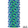 5332C  3j0hC 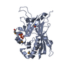 3speC M: atomic model generated by this map C: citing same article ( |
|---|---|
| Similar structure data |

-Links
| EMDB pages | EMDB (EBI/PDBe) / EMDataResource |
|---|
-Map
| File | Download / File: emd_5331.map.gz / Format: CCP4 / Size: 51.5 MB / Type: IMAGE STORED AS FLOATING POINT NUMBER (4 BYTES) | ||||||||||||||||||||||||||||||||||||||||||||||||||||||||||||||||||||
|---|---|---|---|---|---|---|---|---|---|---|---|---|---|---|---|---|---|---|---|---|---|---|---|---|---|---|---|---|---|---|---|---|---|---|---|---|---|---|---|---|---|---|---|---|---|---|---|---|---|---|---|---|---|---|---|---|---|---|---|---|---|---|---|---|---|---|---|---|---|
| Annotation | This is a map of the bacteriophage phiKZ polysheath | ||||||||||||||||||||||||||||||||||||||||||||||||||||||||||||||||||||
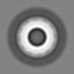
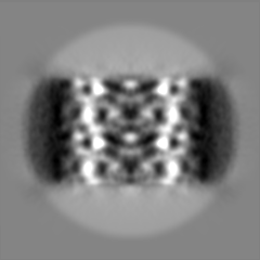
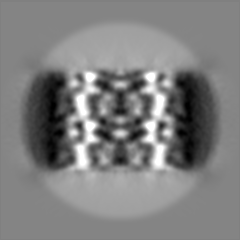
| Projections & slices | Image control
Images are generated by Spider. | ||||||||||||||||||||||||||||||||||||||||||||||||||||||||||||||||||||
| Voxel size | X=Y=Z: 2.49 Å | ||||||||||||||||||||||||||||||||||||||||||||||||||||||||||||||||||||
| Density |
| ||||||||||||||||||||||||||||||||||||||||||||||||||||||||||||||||||||
| Symmetry | Space group: 1 | ||||||||||||||||||||||||||||||||||||||||||||||||||||||||||||||||||||
| Details | EMDB XML:
CCP4 map header:
| ||||||||||||||||||||||||||||||||||||||||||||||||||||||||||||||||||||
 Z (Sec.)
 Y (Row.)
 X (Col.)


-Supplemental data
- Sample components
-Entire : Bacteriophage phiKZ polysheath
| Entire | Name: Bacteriophage phiKZ polysheath |
|---|---|
| Components |
|
-Supramolecule #1000: Bacteriophage phiKZ polysheath
| Supramolecule | Name: Bacteriophage phiKZ polysheath / type: sample / ID: 1000 / Number unique components: 1 |
|---|
-Macromolecule #1: bacteriophage phiKZ polysheaths
| Macromolecule | Name: bacteriophage phiKZ polysheaths / type: protein_or_peptide / ID: 1 / Name.synonym: polysheaths / Recombinant expression: Yes / Database: NCBI |
|---|---|
| Source (natural) | Organism: Pseudomonas phage phiKZ (virus) |
-Experimental details
-Structure determination
| Method | cryo EM |
|---|---|
 Processing Processing | single particle reconstruction |
| Aggregation state | particle |
-Sample preparation
| Vitrification | Cryogen name: ETHANE / Instrument: OTHER |
|---|
- Electron microscopy
| Microscope | FEI/PHILIPS CM200FEG |
|---|---|
| Electron beam | Acceleration voltage: 200 kV / Electron source:  FIELD EMISSION GUN FIELD EMISSION GUN |
| Electron optics | Illumination mode: SPOT SCAN / Imaging mode: BRIGHT FIELD |
| Sample stage | Specimen holder: Eucentric / Specimen holder model: SIDE ENTRY, EUCENTRIC |
-Image processing
| Final reconstruction | Resolution.type: BY AUTHOR / Resolution: 19.0 Å / Resolution method: FSC 0.5 CUT-OFF / Software - Name: SPIDER |
|---|
