 Movie
Movie Controller


+ Open data

- Basic information
| Entry | Database: PDB / ID: 21co | ||||||||||||||||||||||||
|---|---|---|---|---|---|---|---|---|---|---|---|---|---|---|---|---|---|---|---|---|---|---|---|---|---|
| Title | Mfa1 type V pilus from P.gingivalis | ||||||||||||||||||||||||
 Components Components | (Minor fimbrium subunit Mfa1) x 3 | ||||||||||||||||||||||||
 Keywords Keywords | CELL ADHESION / Type V pilus / Mfa1 / Porphyromonas gingivalis / ATCC33277 | ||||||||||||||||||||||||
| Function / homology | pilus shaft / : / Fimbrial subunit protein, C-terminal / Major fimbrial subunit protein type IV, Fimbrillin, C-terminal / outer membrane / cell outer membrane / cell-cell adhesion / Prokaryotic membrane lipoprotein lipid attachment site profile. / Minor fimbrium subunit Mfa1 Function and homology information Function and homology information | ||||||||||||||||||||||||
| Biological species |  Porphyromonas gingivalis ATCC 33277 (bacteria) Porphyromonas gingivalis ATCC 33277 (bacteria) | ||||||||||||||||||||||||
| Method | ELECTRON MICROSCOPY / single particle reconstruction / cryo EM / Resolution: 2.99 Å | ||||||||||||||||||||||||
 Authors Authors | Shibata, S. / Matsunami, H. / Wolf, M. | ||||||||||||||||||||||||
| Funding support |  Japan, 6items Japan, 6items
| ||||||||||||||||||||||||
 Citation Citation | Journal: Commun Biol / Year: 2026 Title: Cryo-EM structure of the native assembled Mfa type V pilus from the periodontal pathogen Porphyromonas gingivalis. Authors: Satoshi Shibata / Hideyuki Matsunami / Kazuhisa Ouhara / Yuri Taniguchi / Makoto Tokoro Schreiber / Alejandro Villar-Brillones / Koji Nakayama / Mikio Shoji / Matthias Wolf /  Abstract: Porphyromonas gingivalis is a primary pathogen causing periodontal disease. The cell has two kinds of type V pili, the Fim pilus and the Mfa pilus, both of which play essential roles in colonization, ...Porphyromonas gingivalis is a primary pathogen causing periodontal disease. The cell has two kinds of type V pili, the Fim pilus and the Mfa pilus, both of which play essential roles in colonization, biofilm formation, and pathogenicity. The functional polymerized structure of the Fim pilus is known, whereas the structure and assembly mechanism of the Mfa pilus remain unclear. Here, we show the structure of the polymerized recombinant Mfa1 stalk pilin determined by cryo-electron microscopy at 3.0 Å resolution. The atomic model of the Mfa1 filament reveals that Mfa1 pilins polymerize by protease-mediated strand exchange and retain a Ca ion in the metal-binding pocket, which modulates immune recognition of the Mfa pilus by human cells. Furthermore, we elucidated the three-dimensional architecture of the streptococcal-binding region on the Mfa pilus. Our results further strengthen evidence that protease-mediated strand exchange is the universal assembly mechanism of type V pili. Our structure of the polymerized Mfa pilus, which represents the functional state on the cell surface, provides targets for antimicrobial drug design to treat periodontal disease and P. gingivalis-related systemic diseases. | ||||||||||||||||||||||||
| History |
|
- Structure visualization
| Structure viewer | Molecule: MolmilJmol/JSmol |
|---|
- Downloads & links
-Download
| PDBx/mmCIF format | 21co.cif.gz | 184 KB | Display | PDBx/mmCIF format |
|---|---|---|---|---|
| PDB format | pdb21co.ent.gz | 143.7 KB | Display | PDB format |
| PDBx/mmJSON format | 21co.json.gz | Tree view | PDBx/mmJSON format | |
| Others |  Other downloads Other downloads |
-Validation report
| Arichive directory | https://data.pdbj.org/pub/pdb/validation_reports/1c/21coftp://data.pdbj.org/pub/pdb/validation_reports/1c/21co | HTTPS FTP |
|---|
-Related structure data
| Related structure data |  67574MC M: map data used to model this data C: citing same article ( |
|---|---|
| Similar structure data |
-Links
 PDBj
PDBj
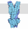
- Assembly
| Deposited unit | 
|
|---|---|
| 1 |
|
-Components
| #1: Protein/peptide | Mass: 2725.075 Da / Num. of mol.: 1 Source method: isolated from a genetically manipulated source Source: (gene. exp.) Porphyromonas gingivalis ATCC 33277 (bacteria)Gene: mfa1, PGN_0287 / Production host:  Escherichia coli BL21(DE3) (bacteria) / References: UniProt: B2RHG1 Escherichia coli BL21(DE3) (bacteria) / References: UniProt: B2RHG1 | ||||
|---|---|---|---|---|---|
| #2: Protein | Mass: 55633.906 Da / Num. of mol.: 1 Source method: isolated from a genetically manipulated source Source: (gene. exp.) Porphyromonas gingivalis ATCC 33277 (bacteria)Gene: mfa1, PGN_0287 / Production host: Escherichia coli BL21(DE3) (bacteria) / References: UniProt: B2RHG1 | ||||
| #3: Protein | Mass: 52926.863 Da / Num. of mol.: 1 Source method: isolated from a genetically manipulated source Source: (gene. exp.) Porphyromonas gingivalis ATCC 33277 (bacteria)Gene: mfa1, PGN_0287 / Production host: Escherichia coli BL21(DE3) (bacteria) / References: UniProt: B2RHG1 | ||||
| #4: Chemical |   Mass: 40.078 Da / Num. of mol.: 2 / Source method: obtained synthetically / Formula: Ca / Feature type: SUBJECT OF INVESTIGATION Mass: 40.078 Da / Num. of mol.: 2 / Source method: obtained synthetically / Formula: Ca / Feature type: SUBJECT OF INVESTIGATIONHas ligand of interest | Y | Has protein modification | N | |
-Experimental details
-Experiment
| Experiment | Method: ELECTRON MICROSCOPY |
|---|---|
| EM experiment | Aggregation state: FILAMENT / 3D reconstruction method: single particle reconstruction |
- Sample preparation
| Component | Name: Mfa1 minor type V pilus / Type: COMPLEX / Entity ID: #1-#3 / Source: RECOMBINANT |
|---|---|
| Molecular weight | Experimental value: NO |
| Source (natural) | Organism: Porphyromonas gingivalis ATCC 33277 (bacteria) |
| Source (recombinant) | Organism: Escherichia coli BL21(DE3) (bacteria) |
| Buffer solution | pH: 7.5 |
| Specimen | Embedding applied: NO / Shadowing applied: NO / Staining applied: NO / Vitrification applied: YES |
| Specimen support | Grid material: COPPER / Grid type: Quantifoil R2/1 |
| Vitrification | Cryogen name: ETHANE-PROPANE |
- Electron microscopy imaging
| Experimental equipment |  Model: Titan Krios / Image courtesy: FEI Company |
|---|---|
| Microscopy | Model: TFS KRIOS |
| Electron gun | Electron source:  FIELD EMISSION GUN / Accelerating voltage: 300 kV / Illumination mode: FLOOD BEAM FIELD EMISSION GUN / Accelerating voltage: 300 kV / Illumination mode: FLOOD BEAM |
| Electron lens | Mode: BRIGHT FIELD / Nominal defocus max: 2250 nm / Nominal defocus min: 1500 nm / Alignment procedure: COMA FREE |
| Specimen holder | Cryogen: NITROGEN / Specimen holder model: FEI TITAN KRIOS AUTOGRID HOLDER |
| Image recording | Electron dose: 32 e/Å2 / Detector mode: INTEGRATING / Film or detector model: FEI FALCON III (4k x 4k) |
- Processing
| EM software |
| |||||||||
|---|---|---|---|---|---|---|---|---|---|---|
| CTF correction | Type: PHASE FLIPPING AND AMPLITUDE CORRECTION | |||||||||
| 3D reconstruction | Resolution: 2.99 Å / Resolution method: FSC 0.143 CUT-OFF / Num. of particles: 267383 / Algorithm: FOURIER SPACE / Symmetry type: POINT |
