 ムービー
ムービー コントローラー


+ データを開く

- 基本情報
基本情報
| 登録情報 | 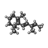 データベース: PDB化学物質要素 / ID: LSR データベース: PDB化学物質要素 / ID: LSR |
|---|---|
| 名称 | 名称: |
-Chemical information
| 組成 |  | ||||
|---|---|---|---|---|---|
| その他 | タイプ: NON-POLYMER / PDB分類: HETAIN / 3文字コード: LSR / 理論座標の詳細: Corina / モデル座標のPDB-ID: 3CR6 | ||||
| 履歴 |
| ||||
 外部リンク 外部リンク | UniChem / ChemSpider / DrugBank / PubChem / SureChEMBL / Wikipedia search / Google search |
- 構造の表示
構造の表示
| 構造ビューア | 分子:  MolmilJmol/JSmol MolmilJmol/JSmol |
|---|
-詳細
-SMILES
| ACDLabs 10.04 | | CACTVS 3.341 | OpenEye OEToolkits 1.5.0 | |
|---|
-SMILES CANONICAL
| CACTVS 3.341 | | OpenEye OEToolkits 1.5.0 | |
|---|
-InChI
| InChI 1.03 |
|---|
-InChIKey
| InChI 1.03 |
|---|
-SYSTEMATIC NAME
| ACDLabs 10.04 | | OpenEye OEToolkits 1.5.0 | |
|---|
-PDBエントリ
全4件を表示しています

PDB-3cr6: 
Crystal Structure of the R132K:R111L:A32E Mutant of Cellular Retinoic Acid Binding Protein Type II Complexed with C15-aldehyde (a retinal analog) at 1.22 Angstrom resolution.

PDB-3f8a:
Crystal Structure of the R132K:R111L:L121E:R59W Mutant of Cellular Retinoic Acid-Binding Protein Type II Complexed with C15-aldehyde (a retinal analog) at 1.95 Angstrom resolution.

PDB-3f9d:
Crystal structure of the R132K:R111L:T54E mutant of cellular retinoic acid-binding protein II complexed with C15-aldehyde (a retinal analog) at 2.00 angstrom resolution

PDB-3fa6:
Crystal structure of the R132K:Y134F:R111L:L121D:T54V mutant of cellular retinoic acid-binding protein II complexed with C15-aldehyde (a retinal analog) at 1.54 angstrom resolution
