 Movie
Movie Controller

[English] 日本語
 Yorodumi
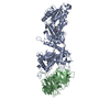
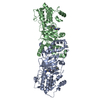
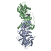
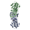
- EMDB-9357: Outer Membrane Cytochrome S Filament from Geobacter Sulfurreducens -
+ Open data

- Basic information
| Entry | Database: EMDB / ID: EMD-9357 | |||||||||
|---|---|---|---|---|---|---|---|---|---|---|
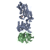
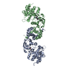
| Title | Outer Membrane Cytochrome S Filament from Geobacter Sulfurreducens | |||||||||
 Map data Map data | ||||||||||
 Sample Sample |
| |||||||||
 Keywords Keywords | Conductive / filament / nanowire / SIX-HEME MULTIHEME C-TYPE CYTOCHROME / PROTEIN FIBRIL | |||||||||
| Function / homology | : / Multiheme cytochrome superfamily / anaerobic respiration / cell outer membrane / electron transfer activity / cell surface / metal ion binding / C-type cytochrome OmcS Function and homology information Function and homology information | |||||||||
| Biological species |  Geobacter sulfurreducens (bacteria) / Geobacter sulfurreducens (strain ATCC 51573 / DSM 12127 / PCA) (bacteria) Geobacter sulfurreducens (bacteria) / Geobacter sulfurreducens (strain ATCC 51573 / DSM 12127 / PCA) (bacteria) | |||||||||
| Method | helical reconstruction / cryo EM / Resolution: 3.4 Å | |||||||||
 Authors Authors | Filman DJ / Marino SF | |||||||||
| Funding support |  United States, 1 items United States, 1 items
| |||||||||
 Citation Citation | Journal: Commun Biol / Year: 2019 Title: Cryo-EM reveals the structural basis of long-range electron transport in a cytochrome-based bacterial nanowire. Authors: David J Filman / Stephen F Marino / Joy E Ward / Lu Yang / Zoltán Mester / Esther Bullitt / Derek R Lovley / Mike Strauss /   Abstract: Electrically conductive pili from species, termed bacterial nanowires, are intensely studied for their biological significance and potential in the development of new materials. Using cryo-electron ...Electrically conductive pili from species, termed bacterial nanowires, are intensely studied for their biological significance and potential in the development of new materials. Using cryo-electron microscopy, we have characterized nanowires from conductive pili preparations that are composed solely of head-to-tail stacked monomers of the six-heme C-type cytochrome OmcS. The unique fold of OmcS - closely wrapped around a continuous stack of hemes that can serve as an uninterrupted path for electron transport - generates a scaffold that supports the unbranched chain of hemes along the central axis of the filament. We present here, at 3.4 Å resolution, the structure of this cytochrome-based filament and discuss its possible role in long-range biological electron transport. | |||||||||
| History |
|
- Structure visualization
| Movie |
Movie viewer |
|---|---|
| Structure viewer | EM map: SurfViewMolmilJmol/JSmol |
| Supplemental images |

- Downloads & links
-EMDB archive
| Map data | emd_9357.map.gz | 944.9 KB | EMDB map data format | |
|---|---|---|---|---|
| Header (meta data) | emd-9357-v30.xmlemd-9357.xml | 11.5 KB 11.5 KB | Display Display | EMDB header |
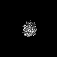
| Images | 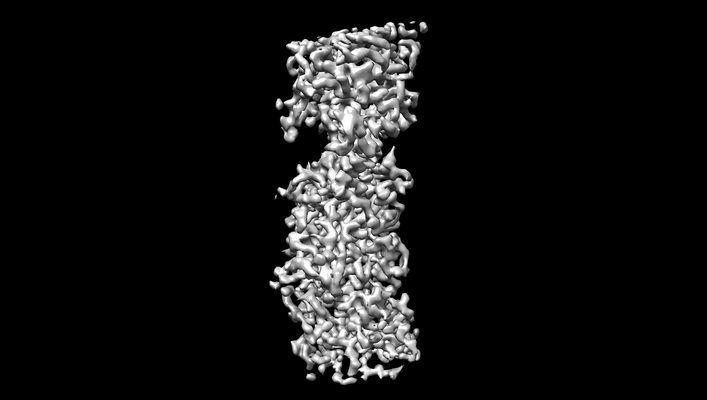 emd_9357.png emd_9357.png | 42.6 KB | ||
| Filedesc metadata | emd-9357.cif.gz | 6 KB | ||
| Archive directory |  http://ftp.pdbj.org/pub/emdb/structures/EMD-9357ftp://ftp.pdbj.org/pub/emdb/structures/EMD-9357 http://ftp.pdbj.org/pub/emdb/structures/EMD-9357ftp://ftp.pdbj.org/pub/emdb/structures/EMD-9357 | HTTPS FTP |
-Related structure data
| Related structure data |  6nefMC M: atomic model generated by this map C: citing same article ( |
|---|---|
| Similar structure data |


-Links
| EMDB pages | EMDB (EBI/PDBe) / EMDataResource |
|---|---|
| Related items in Molecule of the Month |

-Map
| File | Download / File: emd_9357.map.gz / Format: CCP4 / Size: 30.5 MB / Type: IMAGE STORED AS FLOATING POINT NUMBER (4 BYTES) | ||||||||||||||||||||||||||||||||||||||||||||||||||||||||||||||||||||
|---|---|---|---|---|---|---|---|---|---|---|---|---|---|---|---|---|---|---|---|---|---|---|---|---|---|---|---|---|---|---|---|---|---|---|---|---|---|---|---|---|---|---|---|---|---|---|---|---|---|---|---|---|---|---|---|---|---|---|---|---|---|---|---|---|---|---|---|---|---|
| Projections & slices | Image control
Images are generated by Spider. | ||||||||||||||||||||||||||||||||||||||||||||||||||||||||||||||||||||
| Voxel size | X=Y=Z: 1.06 Å | ||||||||||||||||||||||||||||||||||||||||||||||||||||||||||||||||||||
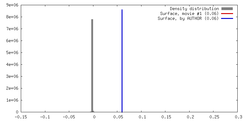
| Density |
| ||||||||||||||||||||||||||||||||||||||||||||||||||||||||||||||||||||
| Symmetry | Space group: 1 | ||||||||||||||||||||||||||||||||||||||||||||||||||||||||||||||||||||
| Details | EMDB XML:
CCP4 map header:
| ||||||||||||||||||||||||||||||||||||||||||||||||||||||||||||||||||||
 Y (Sec.)
 X (Row.)
 Z (Col.)


-Supplemental data
- Sample components
-Entire : Outer Membrane Cytochrome S filament as a bacterial nanowire
| Entire | Name: Outer Membrane Cytochrome S filament as a bacterial nanowire |
|---|---|
| Components |
|
-Supramolecule #1: Outer Membrane Cytochrome S filament as a bacterial nanowire
| Supramolecule | Name: Outer Membrane Cytochrome S filament as a bacterial nanowire type: organelle_or_cellular_component / ID: 1 / Parent: 0 / Macromolecule list: #1 |
|---|---|
| Source (natural) | Organism: Geobacter sulfurreducens (bacteria) |
-Macromolecule #1: C-type cytochrome OmcS
| Macromolecule | Name: C-type cytochrome OmcS / type: protein_or_peptide / ID: 1 Details: SIX-HEME MULTIHEME C-TYPE CYTOCHROME, PROTEIN FIBRIL Number of copies: 1 / Enantiomer: LEVO |
|---|---|
| Source (natural) | Organism: Geobacter sulfurreducens (strain ATCC 51573 / DSM 12127 / PCA) (bacteria) Strain: ATCC 51573 / DSM 12127 / PCA |
| Molecular weight | Theoretical: 42.986008 KDa |
| Sequence | String: FHSGGVAECE GCHTMHNSLG GAVMNSATAQ FTTGPMLLQG ATQSSSCLNC HQHAGDTGPS SYHISTAEAD MPAGTAPLQM TPGGDFGWV KKTYTWNVRG LNTSEGERKG HNIVAGDYNY VADTTLTTAP GGTYPANQLH CSSCHDPHGK YRRFVDGSIA T TGLPIKNS ...String: FHSGGVAECE GCHTMHNSLG GAVMNSATAQ FTTGPMLLQG ATQSSSCLNC HQHAGDTGPS SYHISTAEAD MPAGTAPLQM TPGGDFGWV KKTYTWNVRG LNTSEGERKG HNIVAGDYNY VADTTLTTAP GGTYPANQLH CSSCHDPHGK YRRFVDGSIA T TGLPIKNS GSYQNSNDPT AWGAVGAYRI LGGTGYQPKS LSGSYAFANQ VPAAVAPSTY NRTEATTQTR VAYGQGMSEW CA NCHTDIH NSAYPTNLRH PAGNGAKFGA TIAGLYNSYK KSGDLTGTQA SAYLSLAPFE EGTADYTVLK GHAKIDDTAL TGA DATSNV NCLSCHRAHA SGFDSMTRFN LAYEFTTIAD ASGNSIYGTD PNTSSLQGRS VNEMTAAYYG RTADKFAPYQ RALC NKCHA KD UniProtKB: C-type cytochrome OmcS |
-Macromolecule #2: HEME C
| Macromolecule | Name: HEME C / type: ligand / ID: 2 / Number of copies: 6 / Formula: HEC |
|---|---|
| Molecular weight | Theoretical: 618.503 Da |
| Chemical component information |  ChemComp-HEC: |
-Macromolecule #3: MAGNESIUM ION
| Macromolecule | Name: MAGNESIUM ION / type: ligand / ID: 3 / Number of copies: 1 / Formula: MG |
|---|---|
| Molecular weight | Theoretical: 24.305 Da |
-Experimental details
-Structure determination
| Method | cryo EM |
|---|---|
 Processing Processing | helical reconstruction |
| Aggregation state | filament |
-Sample preparation
| Buffer | pH: 7 |
|---|---|
| Vitrification | Cryogen name: ETHANE / Chamber humidity: 100 % / Chamber temperature: 277 K / Instrument: FEI VITROBOT MARK IV |
| Details | Sample contained thick (4nm) and thin (3nm) filaments. This reconstruction is of the thick filaments. |
- Electron microscopy
| Microscope | FEI TITAN KRIOS |
|---|---|
| Image recording | Film or detector model: GATAN K2 QUANTUM (4k x 4k) / Detector mode: COUNTING / Average electron dose: 45.0 e/Å2 |
| Electron beam | Acceleration voltage: 300 kV / Electron source:  FIELD EMISSION GUN FIELD EMISSION GUN |
| Electron optics | Illumination mode: FLOOD BEAM / Imaging mode: BRIGHT FIELD |
| Experimental equipment |  Model: Titan Krios / Image courtesy: FEI Company |
-Image processing
| Final reconstruction | Applied symmetry - Helical parameters - Δz: 47.5 Å Applied symmetry - Helical parameters - Δ&Phi: 83.01 ° Applied symmetry - Helical parameters - Axial symmetry: C1 (asymmetric) Resolution.type: BY AUTHOR / Resolution: 3.4 Å / Resolution method: FSC 0.143 CUT-OFF / Number images used: 462964 |
|---|---|
| Startup model | Type of model: NONE |
| Final angle assignment | Type: NOT APPLICABLE |
-Atomic model buiding 1
| Details | Atomic model was built de novo visually and repeatedly rebuilt visually using Coot and SPDBV. After each round of manual rebuilding, the parameters of the atomic model underwent stereochemically restrained reciprocal space refinement, using either Refmac5 or Phenix.autobuild or both, using as a reference the amplitudes and phases of the Fourier transform of a portion of the cryoEM reconstruction. For convenience, a single-subunit model was initially built to fit an approximation of the reference map in space group P4(3), which was subsequently replaced by a three-subunit model, using the authentic helical parameters of the map. one of the two axial ligands for HEM 505 is ne2 of his 41 from a neighboring helical-symmetry-related protein chain. |
|---|---|
| Refinement | Space: RECIPROCAL / Protocol: OTHER / Target criteria: maximum likelihood with phases |
| Output model | PDB-6nef: |
