Movie
Movie Controller

[English] 日本語
 Yorodumi
- EMDB-8176: Cryo-EM structure of an ErmBL-stalled ribosome in complex with P-... -
+ Open data

- Basic information
| Entry | Database: EMDB / ID: EMD-8176 | |||||||||
|---|---|---|---|---|---|---|---|---|---|---|
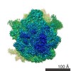
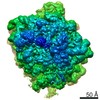
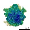
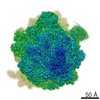
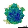
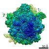
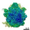
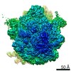
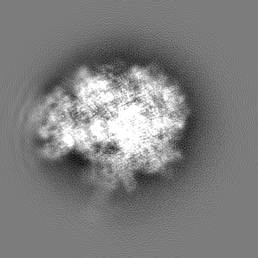
| Title | Cryo-EM structure of an ErmBL-stalled ribosome in complex with P-, and E-tRNA | |||||||||
 Map data Map data | None | |||||||||
 Sample Sample |
| |||||||||
 Keywords Keywords | Ribosome / ErmBL / Stalling / Translation / Erythromycin | |||||||||
| Function / homology |  Function and homology information Function and homology informationstringent response / transcription antitermination factor activity, RNA binding / ornithine decarboxylase inhibitor activity / misfolded RNA binding / Group I intron splicing / RNA folding / translational termination / transcriptional attenuation / endoribonuclease inhibitor activity / positive regulation of ribosome biogenesis ...stringent response / transcription antitermination factor activity, RNA binding / ornithine decarboxylase inhibitor activity / misfolded RNA binding / Group I intron splicing / RNA folding / translational termination / transcriptional attenuation / endoribonuclease inhibitor activity / positive regulation of ribosome biogenesis / RNA-binding transcription regulator activity / four-way junction DNA binding / negative regulation of cytoplasmic translation / DnaA-L2 complex / regulation of mRNA stability / translation repressor activity / negative regulation of translational initiation / negative regulation of DNA-templated DNA replication initiation / mRNA regulatory element binding translation repressor activity / regulation of DNA-templated transcription elongation / positive regulation of RNA splicing / transcription elongation factor complex / response to reactive oxygen species / cytosolic ribosome assembly / ribosome assembly / assembly of large subunit precursor of preribosome / transcription antitermination / DNA endonuclease activity / regulation of cell growth / DNA-templated transcription termination / response to radiation / maintenance of translational fidelity / mRNA 5'-UTR binding / regulation of translation / large ribosomal subunit / transferase activity / ribosomal small subunit assembly / ribosome biogenesis / ribosome binding / ribosomal small subunit biogenesis / 5S rRNA binding / small ribosomal subunit / ribosomal large subunit assembly / small ribosomal subunit rRNA binding / cytosolic small ribosomal subunit / large ribosomal subunit rRNA binding / cytosolic large ribosomal subunit / cytoplasmic translation / tRNA binding / negative regulation of translation / rRNA binding / structural constituent of ribosome / ribosome / translation / response to antibiotic / negative regulation of DNA-templated transcription / hydrolase activity / mRNA binding / DNA binding / RNA binding / zinc ion binding / membrane / cytoplasm / cytosol Similarity search - Function | |||||||||
| Biological species |  | |||||||||
| Method | single particle reconstruction / cryo EM / Resolution: 3.6 Å | |||||||||
 Authors Authors | Arenz S / Bock LV | |||||||||
 Citation Citation | Journal: Nat Commun / Year: 2016 Title: A combined cryo-EM and molecular dynamics approach reveals the mechanism of ErmBL-mediated translation arrest. Authors: Stefan Arenz / Lars V Bock / Michael Graf / C Axel Innis / Roland Beckmann / Helmut Grubmüller / Andrea C Vaiana / Daniel N Wilson /   Abstract: Nascent polypeptides can induce ribosome stalling, regulating downstream genes. Stalling of ErmBL peptide translation in the presence of the macrolide antibiotic erythromycin leads to resistance in ...Nascent polypeptides can induce ribosome stalling, regulating downstream genes. Stalling of ErmBL peptide translation in the presence of the macrolide antibiotic erythromycin leads to resistance in Streptococcus sanguis. To reveal this stalling mechanism we obtained 3.6-Å-resolution cryo-EM structures of ErmBL-stalled ribosomes with erythromycin. The nascent peptide adopts an unusual conformation with the C-terminal Asp10 side chain in a previously unseen rotated position. Together with molecular dynamics simulations, the structures indicate that peptide-bond formation is inhibited by displacement of the peptidyl-tRNA A76 ribose from its canonical position, and by non-productive interactions of the A-tRNA Lys11 side chain with the A-site crevice. These two effects combine to perturb peptide-bond formation by increasing the distance between the attacking Lys11 amine and the Asp10 carbonyl carbon. The interplay between drug, peptide and ribosome uncovered here also provides insight into the fundamental mechanism of peptide-bond formation. | |||||||||
| History |
|
- Structure visualization
| Movie |
Movie viewer |
|---|---|
| Structure viewer | EM map: SurfViewMolmilJmol/JSmol |
| Supplemental images |
- Downloads & links
-EMDB archive
| Map data | emd_8176.map.gz | 176.3 MB | EMDB map data format | |
|---|---|---|---|---|
| Header (meta data) | emd-8176-v30.xmlemd-8176.xml | 69.5 KB 69.5 KB | Display Display | EMDB header |
| Images |  emd_8176.png emd_8176.png | 38.3 KB | ||
| Filedesc metadata | emd-8176.cif.gz | 14.1 KB | ||
| Archive directory |  http://ftp.pdbj.org/pub/emdb/structures/EMD-8176ftp://ftp.pdbj.org/pub/emdb/structures/EMD-8176 http://ftp.pdbj.org/pub/emdb/structures/EMD-8176ftp://ftp.pdbj.org/pub/emdb/structures/EMD-8176 | HTTPS FTP |
-Related structure data
| Related structure data |  5ju8MC  8175C 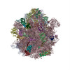 5jteC M: atomic model generated by this map C: citing same article ( |
|---|---|
| Similar structure data |


-Links
| EMDB pages | EMDB (EBI/PDBe) / EMDataResource |
|---|---|
| Related items in Molecule of the Month |


-Map
| File | Download / File: emd_8176.map.gz / Format: CCP4 / Size: 190.1 MB / Type: IMAGE STORED AS FLOATING POINT NUMBER (4 BYTES) | ||||||||||||||||||||||||||||||||||||||||||||||||||||||||||||||||||||
|---|---|---|---|---|---|---|---|---|---|---|---|---|---|---|---|---|---|---|---|---|---|---|---|---|---|---|---|---|---|---|---|---|---|---|---|---|---|---|---|---|---|---|---|---|---|---|---|---|---|---|---|---|---|---|---|---|---|---|---|---|---|---|---|---|---|---|---|---|---|
| Annotation | None | ||||||||||||||||||||||||||||||||||||||||||||||||||||||||||||||||||||
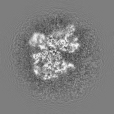
| Projections & slices | Image control
Images are generated by Spider. | ||||||||||||||||||||||||||||||||||||||||||||||||||||||||||||||||||||
| Voxel size | X=Y=Z: 1.108 Å | ||||||||||||||||||||||||||||||||||||||||||||||||||||||||||||||||||||
| Density |
| ||||||||||||||||||||||||||||||||||||||||||||||||||||||||||||||||||||
| Symmetry | Space group: 1 | ||||||||||||||||||||||||||||||||||||||||||||||||||||||||||||||||||||
| Details | EMDB XML:
CCP4 map header:
| ||||||||||||||||||||||||||||||||||||||||||||||||||||||||||||||||||||
 Z (Sec.)
 Y (Row.)
 X (Col.)


-Supplemental data
- Sample components
+Entire : ErmBL-stalled E.coli 70S ribosome
+Supramolecule #1: ErmBL-stalled E.coli 70S ribosome
+Macromolecule #1: 16S ribosomal RNA
+Macromolecule #22: mRNA
+Macromolecule #23: P-site tRNA Aspartate
+Macromolecule #24: E-site tRNA Valine
+Macromolecule #25: 23S ribosomal RNA
+Macromolecule #26: 5S ribosomal RNA
+Macromolecule #2: 30S ribosomal protein S2
+Macromolecule #3: 30S ribosomal protein S3
+Macromolecule #4: 30S ribosomal protein S4
+Macromolecule #5: 30S ribosomal protein S5
+Macromolecule #6: 30S ribosomal protein S6
+Macromolecule #7: 30S ribosomal protein S7
+Macromolecule #8: 30S ribosomal protein S8
+Macromolecule #9: 30S ribosomal protein S9
+Macromolecule #10: 30S ribosomal protein S10
+Macromolecule #11: 30S ribosomal protein S11
+Macromolecule #12: 30S ribosomal protein S12
+Macromolecule #13: 30S ribosomal protein S13
+Macromolecule #14: 30S ribosomal protein S14
+Macromolecule #15: 30S ribosomal protein S15
+Macromolecule #16: 30S ribosomal protein S16
+Macromolecule #17: 30S ribosomal protein S17
+Macromolecule #18: 30S ribosomal protein S18
+Macromolecule #19: 30S ribosomal protein S19
+Macromolecule #20: 30S ribosomal protein S20
+Macromolecule #21: 30S ribosomal protein S21
+Macromolecule #27: 50S ribosomal protein L2
+Macromolecule #28: 50S ribosomal protein L3
+Macromolecule #29: 50S ribosomal protein L4
+Macromolecule #30: 50S ribosomal protein L5
+Macromolecule #31: 50S ribosomal protein L6
+Macromolecule #32: 50S ribosomal protein L9
+Macromolecule #33: 50S ribosomal protein L11
+Macromolecule #34: 50S ribosomal protein L13
+Macromolecule #35: 50S ribosomal protein L14
+Macromolecule #36: 50S ribosomal protein L15
+Macromolecule #37: 50S ribosomal protein L16
+Macromolecule #38: 50S ribosomal protein L17
+Macromolecule #39: 50S ribosomal protein L18
+Macromolecule #40: 50S ribosomal protein L19
+Macromolecule #41: 50S ribosomal protein L20
+Macromolecule #42: 50S ribosomal protein L21
+Macromolecule #43: 50S ribosomal protein L22
+Macromolecule #44: 50S ribosomal protein L23
+Macromolecule #45: 50S ribosomal protein L24
+Macromolecule #46: 50S ribosomal protein L25
+Macromolecule #47: 50S ribosomal protein L27
+Macromolecule #48: 50S ribosomal protein L28
+Macromolecule #49: 50S ribosomal protein L29
+Macromolecule #50: 50S ribosomal protein L30
+Macromolecule #51: 50S ribosomal protein L32
+Macromolecule #52: 50S ribosomal protein L33
+Macromolecule #53: 50S ribosomal protein L34
+Macromolecule #54: 50S ribosomal protein L35
+Macromolecule #55: 50S ribosomal protein L36
+Macromolecule #56: ErmBL
+Macromolecule #57: ERYTHROMYCIN A
-Experimental details
-Structure determination
| Method | cryo EM |
|---|---|
 Processing Processing | single particle reconstruction |
| Aggregation state | particle |
-Sample preparation
| Buffer | pH: 7.4 |
|---|---|
| Sugar embedding | Material: vitreous ice |
| Grid | Model: Quantifoil R3/3 / Material: COPPER / Support film - Material: CARBON / Support film - topology: HOLEY |
| Vitrification | Cryogen name: ETHANE |
- Electron microscopy
| Microscope | FEI TITAN KRIOS |
|---|---|
| Image recording | Film or detector model: FEI FALCON II (4k x 4k) / Detector mode: INTEGRATING / Average electron dose: 4.0 e/Å2 |
| Electron beam | Acceleration voltage: 300 kV / Electron source:  FIELD EMISSION GUN FIELD EMISSION GUN |
| Electron optics | Illumination mode: SPOT SCAN / Imaging mode: BRIGHT FIELD / Nominal defocus max: 1.2 µm / Nominal defocus min: 0.7000000000000001 µm |
| Experimental equipment |  Model: Titan Krios / Image courtesy: FEI Company |
-Image processing
| Startup model | Type of model: INSILICO MODEL |
|---|---|
| Final reconstruction | Resolution.type: BY AUTHOR / Resolution: 3.6 Å / Resolution method: FSC 0.143 CUT-OFF Details: Due to image processing in the absence of spatial frequencies higher than 8 angstroms, the FSC value of 0.143 was used for average resolution determination. Number images used: 85393 |
| Initial angle assignment | Type: PROJECTION MATCHING / Software - Name: SPIDER |
| Final angle assignment | Type: PROJECTION MATCHING / Software - Name: SPIDER |