ATP synthase, F0 complex, subunit H / ATP synthase complex subunit h / ATP synthase, F0 complex, subunit J / ATP synthase protein 8, fungal type / ATP synthase, F0 complex, subunit F, mitochondria, fungi / ATP synthase j chain / Fungal ATP synthase protein 8 (A6L) / Mitochondrial F1-F0 ATP synthase subunit F of fungi / : / Fungal epsilon subunit of F1F0-ATP synthase C-terminal domain ...ATP synthase, F0 complex, subunit H / ATP synthase complex subunit h / ATP synthase, F0 complex, subunit J / ATP synthase protein 8, fungal type / ATP synthase, F0 complex, subunit F, mitochondria, fungi / ATP synthase j chain / Fungal ATP synthase protein 8 (A6L) / Mitochondrial F1-F0 ATP synthase subunit F of fungi / : / Fungal epsilon subunit of F1F0-ATP synthase C-terminal domain / ATP synthase, F0 complex, subunit B/MI25 / ATP synthase, F0 complex, subunit B / Mitochondrial ATP synthase B chain precursor (ATP-synt_B) / ATP synthase, F0 complex, subunit D, mitochondrial / ATP synthase D chain, mitochondrial (ATP5H) / ATP synthase, F0 complex, subunit D superfamily, mitochondrial / ATP synthase, F0 complex, subunit A, bacterial/mitochondria / ATP synthase, F1 complex, epsilon subunit, mitochondrial / ATP synthase, F1 complex, epsilon subunit superfamily, mitochondrial / Mitochondrial ATP synthase epsilon chain / ATPase, OSCP/delta subunit, conserved site / ATP synthase delta (OSCP) subunit signature. / F1F0 ATP synthase OSCP/delta subunit, N-terminal domain superfamily / ATP synthase, F0 complex, subunit A / ATP synthase, F0 complex, subunit A, active site / ATP synthase, F0 complex, subunit A superfamily / ATP synthase A chain / ATP synthase a subunit signature. / ATPase, OSCP/delta subunit / ATP synthase delta (OSCP) subunit / ATP synthase, F1 complex, delta/epsilon subunit / ATP synthase, F1 complex, delta/epsilon subunit, N-terminal / F0F1 ATP synthase delta/epsilon subunit, N-terminal / ATP synthase, Delta/Epsilon chain, beta-sandwich domain / ATP synthase, F0 complex, subunit C / F1F0 ATP synthase subunit C superfamily / ATP synthase, F0 complex, subunit C, DCCD-binding site / ATP synthase c subunit signature. / ATP synthase, F1 complex, gamma subunit conserved site / ATP synthase gamma subunit signature. / ATP synthase, F1 complex, beta subunit / ATP synthase, alpha subunit, C-terminal domain superfamily / : / ATP synthase, F1 complex, gamma subunit / ATP synthase, F1 complex, gamma subunit superfamily / ATP synthase / ATP synthase, alpha subunit, C-terminal / ATP synthase, F1 complex, alpha subunit / ATP synthase, F1 complex, alpha subunit nucleotide-binding domain / ATP synthase alpha/beta chain, C terminal domain / C-terminal domain of V and A type ATP synthase / V-ATPase proteolipid subunit C-like domain / F/V-ATP synthase subunit C superfamily / ATP synthase subunit C / ATPase, F1/V1 complex, beta/alpha subunit, C-terminal / ATP synthase subunit alpha, N-terminal domain-like superfamily / ATPase, F1/V1/A1 complex, alpha/beta subunit, N-terminal domain superfamily / ATPase, F1/V1/A1 complex, alpha/beta subunit, N-terminal domain / ATP synthase alpha/beta family, beta-barrel domain / ATPase, alpha/beta subunit, nucleotide-binding domain, active site / ATP synthase alpha and beta subunits signature. / ATPase, F1/V1/A1 complex, alpha/beta subunit, nucleotide-binding domain / ATP synthase alpha/beta family, nucleotide-binding domain / ATPases associated with a variety of cellular activities / AAA+ ATPase domain / P-loop containing nucleoside triphosphate hydrolase 類似検索 - ドメイン・相同性
ATP synthase subunit beta, mitochondrial / ATP synthase subunit a / ATP synthase protein 8 / ATP synthase subunit 4, mitochondrial / ATP synthase subunit alpha, mitochondrial / ATP synthase subunit 5, mitochondrial / ATP synthase subunit epsilon, mitochondrial / ATP synthase subunit d, mitochondrial / ATP synthase subunit gamma, mitochondrial / ATP synthase subunit 9, mitochondrial ...ATP synthase subunit beta, mitochondrial / ATP synthase subunit a / ATP synthase protein 8 / ATP synthase subunit 4, mitochondrial / ATP synthase subunit alpha, mitochondrial / ATP synthase subunit 5, mitochondrial / ATP synthase subunit epsilon, mitochondrial / ATP synthase subunit d, mitochondrial / ATP synthase subunit gamma, mitochondrial / ATP synthase subunit 9, mitochondrial / ATP synthase subunit J, mitochondrial / ATP synthase subunit f, mitochondrial / ATP synthase subunit delta, mitochondrial / ATP synthase subunit H, mitochondrial 類似検索 - 構成要素
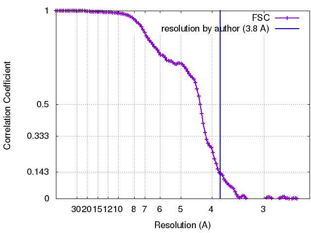
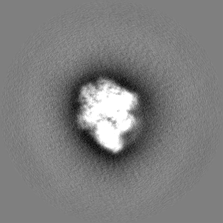
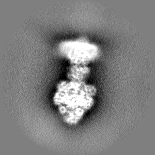
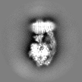
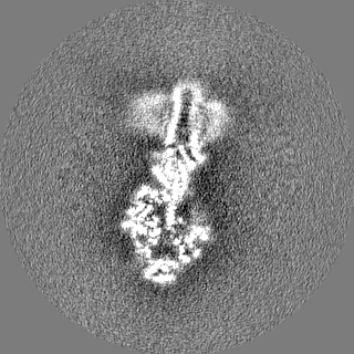
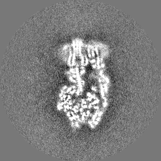
ジャーナル: Science / 年: 2018 タイトル: High-resolution cryo-EM analysis of the yeast ATP synthase in a lipid membrane. 著者: Anurag P Srivastava / Min Luo / Wenchang Zhou / Jindrich Symersky / Dongyang Bai / Melissa G Chambers / José D Faraldo-Gómez / Maofu Liao / David M Mueller / 要旨: Mitochondrial adenosine triphosphate (ATP) synthase comprises a membrane embedded F motor that rotates to drive ATP synthesis in the F subunit. We used single-particle cryo-electron microscopy (cryo- ...Mitochondrial adenosine triphosphate (ATP) synthase comprises a membrane embedded F motor that rotates to drive ATP synthesis in the F subunit. We used single-particle cryo-electron microscopy (cryo-EM) to obtain structures of the full complex in a lipid bilayer in the absence or presence of the inhibitor oligomycin at 3.6- and 3.8-angstrom resolution, respectively. To limit conformational heterogeneity, we locked the rotor in a single conformation by fusing the F6 subunit of the stator with the δ subunit of the rotor. Assembly of the enzyme with the F6-δ fusion caused a twisting of the rotor and a 9° rotation of the F c-ring in the direction of ATP synthesis, relative to the structure of isolated F Our cryo-EM structures show how F and F are coupled, give insight into the proton translocation pathway, and show how oligomycin blocks ATP synthesis.
 ムービー
ムービー コントローラー


 データを開く
 基本情報
基本情報 マップデータ
 試料
試料 キーワード
キーワード 機能・相同性情報
 データ登録者
 米国, 1件
  引用
引用 構造の表示
構造の表示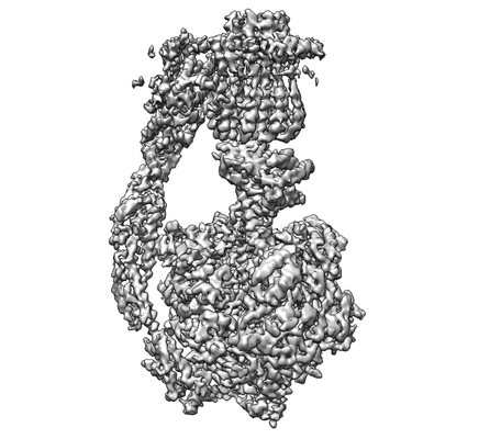
 ダウンロードとリンク
 emd_7546.png
 http://ftp.pdbj.org/pub/emdb/structures/EMD-7546


 Z (Sec.)
 Y (Row.)
 X (Col.)


 試料の構成要素

 解析
解析 電子顕微鏡法
 FIELD EMISSION GUN