 Movie
Movie Controller

[English] 日本語
 Yorodumi
- EMDB-70965: CryoEM structure of Ggust-coupled TAS2R43 with aristolochic acid I -
+ Open data

- Basic information
| Entry |  | |||||||||
|---|---|---|---|---|---|---|---|---|---|---|
| Title | CryoEM structure of Ggust-coupled TAS2R43 with aristolochic acid I | |||||||||
 Map data Map data | ||||||||||
 Sample Sample |
| |||||||||
 Keywords Keywords | GPCR / SIGNALING PROTEIN | |||||||||
| Function / homology |  Function and homology information Function and homology informationbitter taste receptor activity / taste receptor activity / detection of light stimulus involved in visual perception / sensory perception of sweet taste / detection of chemical stimulus involved in sensory perception of bitter taste / motile cilium / Class C/3 (Metabotropic glutamate/pheromone receptors) / ciliary membrane / phototransduction, visible light / axoneme ...bitter taste receptor activity / taste receptor activity / detection of light stimulus involved in visual perception / sensory perception of sweet taste / detection of chemical stimulus involved in sensory perception of bitter taste / motile cilium / Class C/3 (Metabotropic glutamate/pheromone receptors) / ciliary membrane / phototransduction, visible light / axoneme / Adenylate cyclase inhibitory pathway / acrosomal vesicle / response to nicotine / G protein-coupled receptor binding / G protein-coupled receptor activity / G-protein beta/gamma-subunit complex binding / adenylate cyclase-modulating G protein-coupled receptor signaling pathway / Olfactory Signaling Pathway / Activation of the phototransduction cascade / G protein-coupled acetylcholine receptor signaling pathway / G beta:gamma signalling through PLC beta / Presynaptic function of Kainate receptors / Thromboxane signalling through TP receptor / Activation of G protein gated Potassium channels / Inhibition of voltage gated Ca2+ channels via Gbeta/gamma subunits / G-protein activation / Glucagon signaling in metabolic regulation / Prostacyclin signalling through prostacyclin receptor / G beta:gamma signalling through CDC42 / Synthesis, secretion, and inactivation of Glucagon-like Peptide-1 (GLP-1) / G beta:gamma signalling through BTK / photoreceptor disc membrane / ADP signalling through P2Y purinoceptor 12 / Glucagon-type ligand receptors / Sensory perception of sweet, bitter, and umami (glutamate) taste / Adrenaline,noradrenaline inhibits insulin secretion / Vasopressin regulates renal water homeostasis via Aquaporins / Glucagon-like Peptide-1 (GLP1) regulates insulin secretion / G alpha (z) signalling events / cellular response to catecholamine stimulus / ADP signalling through P2Y purinoceptor 1 / ADORA2B mediated anti-inflammatory cytokines production / G beta:gamma signalling through PI3Kgamma / adenylate cyclase-activating dopamine receptor signaling pathway / Cooperation of PDCL (PhLP1) and TRiC/CCT in G-protein beta folding / GPER1 signaling / cellular response to prostaglandin E stimulus / heterotrimeric G-protein complex / G alpha (12/13) signalling events / Inactivation, recovery and regulation of the phototransduction cascade / G-protein beta-subunit binding / extracellular vesicle / sensory perception of taste / Thrombin signalling through proteinase activated receptors (PARs) / signaling receptor complex adaptor activity / retina development in camera-type eye / GTPase binding / fibroblast proliferation / Ca2+ pathway / High laminar flow shear stress activates signaling by PIEZO1 and PECAM1:CDH5:KDR in endothelial cells / G alpha (i) signalling events / G alpha (s) signalling events / G alpha (q) signalling events / phospholipase C-activating G protein-coupled receptor signaling pathway / Ras protein signal transduction / Extra-nuclear estrogen signaling / cell population proliferation / apical plasma membrane / G protein-coupled receptor signaling pathway / lysosomal membrane / GTPase activity / synapse / GTP binding / protein-containing complex binding / signal transduction / protein-containing complex / extracellular exosome / membrane / metal ion binding / plasma membrane / cytoplasm / cytosol Similarity search - Function | |||||||||
| Biological species |  Homo sapiens (human) Homo sapiens (human) | |||||||||
| Method | single particle reconstruction / cryo EM / Resolution: 3.0 Å | |||||||||
 Authors Authors | Kim Y / Gumpper RH / Roth BL | |||||||||
| Funding support |  United States, 1 items United States, 1 items
| |||||||||
 Citation Citation | Journal: Nat Struct Mol Biol / Year: 2026 Title: Structural insights into coffee bitter taste perception by TAS2R43 receptor. Authors: Yoojoong Kim / Ryan H Gumpper / Yuxuan Zhuang / Ron O Dror / Bryan L Roth / Abstract: Bitter taste functions as a means of both protection against potentially toxic compounds and savoring bitter tasting foods and beverages. Among the 26 bitter taste receptors, taste receptor type 2 ...Bitter taste functions as a means of both protection against potentially toxic compounds and savoring bitter tasting foods and beverages. Among the 26 bitter taste receptors, taste receptor type 2 member 43 (TAS2R43) has been identified as key for recognizing the bitter taste of coffee. TAS2R43 has also been implicated in many other physiological processes, including the regulation of glucagon-like peptide 1 release from the intestine, bronchodilation, innate immunity and metabolism. Here we report cryo-electron microscopy structures of human TAS2R43 coupled with inhibitory G protein or gustducin (G) stabilized by the potent nephrotoxin and carcinogen aristolochic acid I. Both structures revealed that aristolochic acid I binds in a presumed orthosteric pocket shared with other bitter taste receptor. Further structural, functional and computational studies revealed potential modes for coffee's constituents including caffeine and cafestol, which are bitter tastants from coffee. Lastly, long-timescale molecular dynamics simulations identified potential cryptic allosteric pockets in TAS2R43. These structures could accelerate the search for specific bitter taste ligands that ultimately may be therapeutically useful. | |||||||||
| History |
|
- Structure visualization
| Supplemental images |
|---|

- Downloads & links
-EMDB archive
| Map data | emd_70965.map.gz | 85.5 MB | EMDB map data format | |
|---|---|---|---|---|
| Header (meta data) | emd-70965-v30.xmlemd-70965.xml | 24.7 KB 24.7 KB | Display Display | EMDB header |
| Images |  emd_70965.png emd_70965.png | 95.5 KB | ||
| Filedesc metadata | emd-70965.cif.gz | 7.2 KB | ||
| Others | emd_70965_half_map_1.map.gzemd_70965_half_map_2.map.gz | 84.3 MB 84.3 MB | ||
| Archive directory |  http://ftp.pdbj.org/pub/emdb/structures/EMD-70965ftp://ftp.pdbj.org/pub/emdb/structures/EMD-70965 http://ftp.pdbj.org/pub/emdb/structures/EMD-70965ftp://ftp.pdbj.org/pub/emdb/structures/EMD-70965 | HTTPS FTP |
-Related structure data
| Related structure data |  9oxbMC  9oxaC C: citing same article ( M: atomic model generated by this map |
|---|---|
| Similar structure data |


-Links
| EMDB pages | EMDB (EBI/PDBe) / EMDataResource |
|---|---|
| Related items in Molecule of the Month |


-Map
| File | Download / File: emd_70965.map.gz / Format: CCP4 / Size: 91.1 MB / Type: IMAGE STORED AS FLOATING POINT NUMBER (4 BYTES) | ||||||||||||||||||||||||||||||||||||
|---|---|---|---|---|---|---|---|---|---|---|---|---|---|---|---|---|---|---|---|---|---|---|---|---|---|---|---|---|---|---|---|---|---|---|---|---|---|
| Projections & slices | Image control
Images are generated by Spider. | ||||||||||||||||||||||||||||||||||||
| Voxel size | X=Y=Z: 0.876 Å | ||||||||||||||||||||||||||||||||||||
| Density |
| ||||||||||||||||||||||||||||||||||||
| Symmetry | Space group: 1 | ||||||||||||||||||||||||||||||||||||
| Details | EMDB XML:
|
 Z (Sec.)
 Y (Row.)
 X (Col.)


-Supplemental data
-Half map: #2
| File | emd_70965_half_map_1.map | ||||||||||||
|---|---|---|---|---|---|---|---|---|---|---|---|---|---|
| Projections & Slices |
| ||||||||||||
| Density Histograms |


-Half map: #1
| File | emd_70965_half_map_2.map | ||||||||||||
|---|---|---|---|---|---|---|---|---|---|---|---|---|---|
| Projections & Slices |
| ||||||||||||
| Density Histograms |


- Sample components
-Entire : GPCR complex 1
| Entire | Name: GPCR complex 1 |
|---|---|
| Components |
|
-Supramolecule #1: GPCR complex 1
| Supramolecule | Name: GPCR complex 1 / type: complex / ID: 1 / Parent: 0 / Macromolecule list: #1-#5 |
|---|---|
| Source (natural) | Organism: Homo sapiens (human) |
-Macromolecule #1: Guanine nucleotide-binding protein G(t) subunit alpha-3
| Macromolecule | Name: Guanine nucleotide-binding protein G(t) subunit alpha-3 type: protein_or_peptide / ID: 1 / Number of copies: 1 / Enantiomer: LEVO |
|---|---|
| Source (natural) | Organism: Homo sapiens (human) |
| Molecular weight | Theoretical: 40.339848 KDa |
| Recombinant expression | Organism:   Spodoptera frugiperda (fall armyworm) Spodoptera frugiperda (fall armyworm) |
| Sequence | String: MGSTVSAEDK AAAERSKMID KNLREDAERD ARTVKLLLLG AGESGKATIV KQQMIIHKNG YSEQECMEFK AVIYSNTLQS ILAIVKAMT TLGIDYVNPR SAEDQRQLYA MANTLEDGGM TPQLAEVIKR LWRDPGIQAC FERASEYQLN DSAAYYLNDL D RITASGYV ...String: MGSTVSAEDK AAAERSKMID KNLREDAERD ARTVKLLLLG AGESGKATIV KQQMIIHKNG YSEQECMEFK AVIYSNTLQS ILAIVKAMT TLGIDYVNPR SAEDQRQLYA MANTLEDGGM TPQLAEVIKR LWRDPGIQAC FERASEYQLN DSAAYYLNDL D RITASGYV PNEQDVLHSR VKTTGIIETQ FSFKDLHFRM FDVGAQRSER KKWIHCFEGV TCIIFCAALS AYDMVLVEDE EV NRMHASL KLFDSICNHK YFSDTSIVLF LNKKDIFQEK VTKVHLSICF PEYTGPNTFE DAGNYIKNQF LDLNLKKEDK EIY SHMTCS TDTQNVKFVF DAVTDIIIKE NLKDCGLF UniProtKB: Guanine nucleotide-binding protein G(t) subunit alpha-3 |
-Macromolecule #2: Guanine nucleotide-binding protein G(I)/G(S)/G(T) subunit beta-1
| Macromolecule | Name: Guanine nucleotide-binding protein G(I)/G(S)/G(T) subunit beta-1 type: protein_or_peptide / ID: 2 / Number of copies: 1 / Enantiomer: LEVO |
|---|---|
| Source (natural) | Organism: Homo sapiens (human) |
| Molecular weight | Theoretical: 42.936668 KDa |
| Recombinant expression | Organism: Spodoptera frugiperda (fall armyworm) |
| Sequence | String: MHHHHHHLEV LFQGPGSSGS ELDQLRQEAE QLKNQIRDAR KACADATLSQ ITNNIDPVGR IQMRTRRTLR GHLAKIYAMH WGTDSRLLV SASQDGKLII WDSYTTNKVH AIPLRSSWVM TCAYAPSGNY VACGGLDNIC SIYNLKTREG NVRVSRELAG H TGYLSCCR ...String: MHHHHHHLEV LFQGPGSSGS ELDQLRQEAE QLKNQIRDAR KACADATLSQ ITNNIDPVGR IQMRTRRTLR GHLAKIYAMH WGTDSRLLV SASQDGKLII WDSYTTNKVH AIPLRSSWVM TCAYAPSGNY VACGGLDNIC SIYNLKTREG NVRVSRELAG H TGYLSCCR FLDDNQIVTS SGDTTCALWD IETGQQTTTF TGHTGDVMSL SLAPDTRLFV SGACDASAKL WDVREGMCRQ TF TGHESDI NAICFFPNGN AFATGSDDAT CRLFDLRADQ ELMTYSHDNI ICGITSVSFS KSGRLLLAGY DDFNCNVWDA LKA DRAGVL AGHDNRVSCL GVTDDGMAVA TGSWDSFLKI WNGGSGGGGS GGSSSGGGGS GGGGSGGSSS GGVSGWRLFK KISG GS UniProtKB: Guanine nucleotide-binding protein G(I)/G(S)/G(T) subunit beta-1 |
-Macromolecule #3: Guanine nucleotide-binding protein G(I)/G(S)/G(O) subunit gamma-2
| Macromolecule | Name: Guanine nucleotide-binding protein G(I)/G(S)/G(O) subunit gamma-2 type: protein_or_peptide / ID: 3 / Number of copies: 1 / Enantiomer: LEVO |
|---|---|
| Source (natural) | Organism: Homo sapiens (human) |
| Molecular weight | Theoretical: 7.861143 KDa |
| Recombinant expression | Organism: Spodoptera frugiperda (fall armyworm) |
| Sequence | String: MASNNTASIA QARKLVEQLK MEANIDRIKV SKAAADLMAY CEAHAKEDPL LTPVPASENP FREKKFFCAI L UniProtKB: Guanine nucleotide-binding protein G(I)/G(S)/G(O) subunit gamma-2 |
-Macromolecule #4: scFv16
| Macromolecule | Name: scFv16 / type: protein_or_peptide / ID: 4 / Number of copies: 1 / Enantiomer: LEVO |
|---|---|
| Source (natural) | Organism: Homo sapiens (human) |
| Molecular weight | Theoretical: 28.668922 KDa |
| Recombinant expression | Organism: Spodoptera frugiperda (fall armyworm) |
| Sequence | String: DVQLVESGGG LVQPGGSRKL SCSASGFAFS SFGMHWVRQA PEKGLEWVAY ISSGSGTIYY ADTVKGRFTI SRDDPKNTLF LQMTSLRSE DTAMYYCVRS IYYYGSSPFD FWGQGTTLTV SSGGGGSGGG GSGGGGSDIV MTQATSSVPV TPGESVSISC R SSKSLLHS ...String: DVQLVESGGG LVQPGGSRKL SCSASGFAFS SFGMHWVRQA PEKGLEWVAY ISSGSGTIYY ADTVKGRFTI SRDDPKNTLF LQMTSLRSE DTAMYYCVRS IYYYGSSPFD FWGQGTTLTV SSGGGGSGGG GSGGGGSDIV MTQATSSVPV TPGESVSISC R SSKSLLHS NGNTYLYWFL QRPGQSPQLL IYRMSNLASG VPDRFSGSGS GTAFTLTISR LEAEDVGVYY CMQHLEYPLT FG AGTKLEL KAAALEVLFQ GPHHHHHHHH |
-Macromolecule #5: Taste receptor type 2 member 43
| Macromolecule | Name: Taste receptor type 2 member 43 / type: protein_or_peptide / ID: 5 / Number of copies: 1 / Enantiomer: LEVO |
|---|---|
| Source (natural) | Organism: Homo sapiens (human) |
| Molecular weight | Theoretical: 72.130312 KDa |
| Recombinant expression | Organism: Spodoptera frugiperda (fall armyworm) |
| Sequence | String: DYKDDDDAKL QTMHHHHHHH HHHENLYFQG GTTMADLEDN WETLNDNLKV IEKADNAAQV KDALTKMRAA ALDAQKATPP KLEDKSPDS PEMKDFRHGF DILVGQIDDA LKLANEGKVK EAQAAAEQLK TTRNAYIQKY LGSTLEVLFQ GPMITFLPII F SSLVVVTF ...String: DYKDDDDAKL QTMHHHHHHH HHHENLYFQG GTTMADLEDN WETLNDNLKV IEKADNAAQV KDALTKMRAA ALDAQKATPP KLEDKSPDS PEMKDFRHGF DILVGQIDDA LKLANEGKVK EAQAAAEQLK TTRNAYIQKY LGSTLEVLFQ GPMITFLPII F SSLVVVTF VIGNFANGFI ALVNSIEWFK RQKISFADQI LTALAVSRVG LLWVLLLNWY STVLNPAFNS VEVRTTAYNI WA VINHFSN WLATTLSIFY LLKIANFSNF IFLHLKRRVK SVILVMLLGP LLFLACHLFV INMNEIVRTK EFEGNMTWKI KLK SAMYFS NMTVTMVANL VPFTLTLLSF MLLICSLCKH LKKMQLHGKG SQDPSTKVHI KALQTVISFL LLCAIYFLSI MISV WSFGS LENKPVFMFC KAIRFSYPSI HPFILIWGNK KLKQTFLSVF WQMRYWVKGE KTSSPGSAGS AGSGGSGGGS GGGGS GGSS SGGVFTLEDF VGDWEQTAAY NLDQVLEQGG VSSLLQNLAV SVTPIQRIVR SGENALKIDI HVIIPYEGLS ADQMAQ IEE VFKVVYPVDD HHFKVILPYG TLVIDGVTPN MLNYFGRPYE GIAVFDGKKI TVTGTLWNGN KIIDERLITP DGSMLFR VT INS UniProtKB: Taste receptor type 2 member 43 |
-Macromolecule #6: 8-methoxy-6-nitro-naphtho[1,2-e][1,3]benzodioxole-5-carboxylic acid
| Macromolecule | Name: 8-methoxy-6-nitro-naphtho[1,2-e][1,3]benzodioxole-5-carboxylic acid type: ligand / ID: 6 / Number of copies: 1 / Formula: GOQ |
|---|---|
| Molecular weight | Theoretical: 341.272 Da |
| Chemical component information | 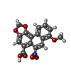 ChemComp-GOQ: |
-Macromolecule #7: water
| Macromolecule | Name: water / type: ligand / ID: 7 / Number of copies: 5 / Formula: HOH |
|---|---|
| Molecular weight | Theoretical: 18.015 Da |
| Chemical component information |  ChemComp-HOH: |
-Experimental details
-Structure determination
| Method | cryo EM |
|---|---|
 Processing Processing | single particle reconstruction |
| Aggregation state | particle |
-Sample preparation
| Concentration | 7 mg/mL |
|---|---|
| Buffer | pH: 7.5 |
| Vitrification | Cryogen name: ETHANE-PROPANE / Chamber humidity: 95 % / Chamber temperature: 277.15 K / Instrument: FEI VITROBOT MARK IV |
- Electron microscopy
| Microscope | FEI TALOS ARCTICA |
|---|---|
| Image recording | Film or detector model: GATAN K3 (6k x 4k) / Average electron dose: 60.0 e/Å2 |
| Electron beam | Acceleration voltage: 200 kV / Electron source:  FIELD EMISSION GUN FIELD EMISSION GUN |
| Electron optics | Illumination mode: FLOOD BEAM / Imaging mode: BRIGHT FIELD / Nominal defocus max: 20.0 µm / Nominal defocus min: 5.0 µm |
| Experimental equipment |  Model: Talos Arctica / Image courtesy: FEI Company |

