Movie
Movie Controller


+ Open data

- Basic information
| Entry |  | |||||||||
|---|---|---|---|---|---|---|---|---|---|---|
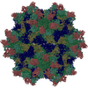
| Title | SIPV1-5E12 Complex | |||||||||
 Map data Map data | ||||||||||
 Sample Sample |
| |||||||||
 Keywords Keywords | Virus / Antibody / Complex | |||||||||
| Function / homology |  Function and homology information Function and homology informationsymbiont-mediated suppression of host cytoplasmic pattern recognition receptor signaling pathway via inhibition of RIG-I activity / symbiont-mediated suppression of host cytoplasmic pattern recognition receptor signaling pathway via inhibition of MDA-5 activity / receptor-mediated endocytosis of virus by host cell / symbiont-mediated suppression of host cytoplasmic pattern recognition receptor signaling pathway via inhibition of MAVS activity / picornain 2A / symbiont-mediated suppression of host mRNA export from nucleus / symbiont genome entry into host cell via pore formation in plasma membrane / picornain 3C / T=pseudo3 icosahedral viral capsid / host cell cytoplasmic vesicle membrane ...symbiont-mediated suppression of host cytoplasmic pattern recognition receptor signaling pathway via inhibition of RIG-I activity / symbiont-mediated suppression of host cytoplasmic pattern recognition receptor signaling pathway via inhibition of MDA-5 activity / receptor-mediated endocytosis of virus by host cell / symbiont-mediated suppression of host cytoplasmic pattern recognition receptor signaling pathway via inhibition of MAVS activity / picornain 2A / symbiont-mediated suppression of host mRNA export from nucleus / symbiont genome entry into host cell via pore formation in plasma membrane / picornain 3C / T=pseudo3 icosahedral viral capsid / host cell cytoplasmic vesicle membrane / ribonucleoside triphosphate phosphatase activity / nucleoside-triphosphate phosphatase / channel activity / monoatomic ion transmembrane transport / DNA replication / RNA helicase activity / symbiont-mediated activation of host autophagy / RNA-directed RNA polymerase / cysteine-type endopeptidase activity / viral RNA genome replication / RNA-directed RNA polymerase activity / virion attachment to host cell / host cell nucleus / DNA-templated transcription / structural molecule activity / proteolysis / RNA binding / zinc ion binding / ATP binding Similarity search - Function | |||||||||
| Biological species |  Human poliovirus 1 strain Sabin / Human poliovirus 1 strain Sabin /  Homo sapiens (human) Homo sapiens (human) | |||||||||
| Method | single particle reconstruction / cryo EM / Resolution: 2.84 Å | |||||||||
 Authors Authors | Waddey BT / Hafenstein SL | |||||||||
| Funding support |  United States, 1 items United States, 1 items
| |||||||||
 Citation Citation | Journal: Nat Commun / Year: 2026 Title: Neutralizing human monoclonal antibodies to poliovirus map to the receptor binding site. Authors: Benjamin T Waddey / Andrew J Charnesky / Julia E Faust / Nadia M DiNunno / Rama Devudu Puligedda / Sung Hyun Cho / Carol M Bator / Steven D Dong / Kutub Mahmood / Konstantin M Chumakov / ...Authors: Benjamin T Waddey / Andrew J Charnesky / Julia E Faust / Nadia M DiNunno / Rama Devudu Puligedda / Sung Hyun Cho / Carol M Bator / Steven D Dong / Kutub Mahmood / Konstantin M Chumakov / Scott K Dessain / Susan L Hafenstein / Abstract: Poliovirus remains a serious threat to human health. Complete eradication of wild-type poliovirus has not yet succeeded, making the development of successful antivirals critical. Microneutralization ...Poliovirus remains a serious threat to human health. Complete eradication of wild-type poliovirus has not yet succeeded, making the development of successful antivirals critical. Microneutralization assays against all three poliovirus serotypes identified a panel of human monoclonal IgGs, which are either serotype-specific or cross-neutralizing. Here, through cryoEM single particle analysis, we solved high resolution structures of four distinct poliovirus-FAb complexes. These antibodies bind to capsids at the circular depression (canyon) surrounding the icosahedral five-fold symmetry axis, which is also the binding site of the poliovirus receptor (PVR). Analysis of these structures confirms overlap of FAb contacts on the viral capsid with those of PVR. For three of the FAbs, the capsid residues are identified that dictate serotype-specific recognition. Contacts for the cross-neutralizing mAb 10D2 are located deep in the capsid canyon. These structural analyses indicate that antibody competition with the receptor likely leads to neutralization of virus particles and inhibition of poliovirus entry into host cells. Thus, the human IgGs studied here may facilitate development of therapeutics for the ongoing efforts in global eradication of poliovirus. | |||||||||
| History |
|
- Structure visualization
| Supplemental images |
|---|

- Downloads & links
-EMDB archive
| Map data | emd_70392.map.gz | 778.6 MB | EMDB map data format | |
|---|---|---|---|---|
| Header (meta data) | emd-70392-v30.xmlemd-70392.xml | 24 KB 24 KB | Display Display | EMDB header |
| FSC (resolution estimation) | emd_70392_fsc.xml | 19.7 KB | Display | FSC data file |
| Images |  emd_70392.png emd_70392.png | 36.2 KB | ||
| Filedesc metadata | emd-70392.cif.gz | 6.6 KB | ||
| Others | emd_70392_half_map_1.map.gzemd_70392_half_map_2.map.gz | 763 MB 762.9 MB | ||
| Archive directory |  http://ftp.pdbj.org/pub/emdb/structures/EMD-70392ftp://ftp.pdbj.org/pub/emdb/structures/EMD-70392 http://ftp.pdbj.org/pub/emdb/structures/EMD-70392ftp://ftp.pdbj.org/pub/emdb/structures/EMD-70392 | HTTPS FTP |
-Related structure data
| Related structure data |  9oeaMC  9oclC  9ocoC  9od3C M: atomic model generated by this map C: citing same article ( |
|---|---|
| Similar structure data |


-Links
| EMDB pages | EMDB (EBI/PDBe) / EMDataResource |
|---|---|
| Related items in Molecule of the Month |


-Map
| File | Download / File: emd_70392.map.gz / Format: CCP4 / Size: 824 MB / Type: IMAGE STORED AS FLOATING POINT NUMBER (4 BYTES) | ||||||||||||||||||||||||||||||||||||
|---|---|---|---|---|---|---|---|---|---|---|---|---|---|---|---|---|---|---|---|---|---|---|---|---|---|---|---|---|---|---|---|---|---|---|---|---|---|
| Projections & slices | Image control
Images are generated by Spider. | ||||||||||||||||||||||||||||||||||||
| Voxel size | X=Y=Z: 1.26 Å | ||||||||||||||||||||||||||||||||||||
| Density |
| ||||||||||||||||||||||||||||||||||||
| Symmetry | Space group: 1 | ||||||||||||||||||||||||||||||||||||
| Details | EMDB XML:
|
 Z (Sec.)
 Y (Row.)
 X (Col.)


-Supplemental data
-Half map: #1
| File | emd_70392_half_map_1.map | ||||||||||||
|---|---|---|---|---|---|---|---|---|---|---|---|---|---|
| Projections & Slices |
| ||||||||||||
| Density Histograms |


-Half map: #2
| File | emd_70392_half_map_2.map | ||||||||||||
|---|---|---|---|---|---|---|---|---|---|---|---|---|---|
| Projections & Slices |
| ||||||||||||
| Density Histograms |


- Sample components
-Entire : SIPV1-5E12 FAb complex
| Entire | Name: SIPV1-5E12 FAb complex |
|---|---|
| Components |
|
-Supramolecule #1: SIPV1-5E12 FAb complex
| Supramolecule | Name: SIPV1-5E12 FAb complex / type: complex / ID: 1 / Parent: 0 / Macromolecule list: #1-#6 |
|---|---|
| Source (natural) | Organism: Human poliovirus 1 strain Sabin |
| Molecular weight | Theoretical: 8 MDa |
-Macromolecule #1: Capsid protein VP1
| Macromolecule | Name: Capsid protein VP1 / type: protein_or_peptide / ID: 1 / Number of copies: 1 / Enantiomer: LEVO |
|---|---|
| Source (natural) | Organism: Human poliovirus 1 strain Sabin |
| Molecular weight | Theoretical: 33.492605 KDa |
| Sequence | String: GLGQMLESMI DNTVRETVGA ATSRDALPNT EASGPAHSKE IPALTAVETG ATNPLVPSDT VQTRHVVQHR SRSESSIESF FARGACVAI ITVDNSASTK NKDKLFTVWK ITYKDTVQLR RKLEFFTYSR FDMEFTFVVT ANFTETNNGH ALNQVYQIMY V PPGAPVPE ...String: GLGQMLESMI DNTVRETVGA ATSRDALPNT EASGPAHSKE IPALTAVETG ATNPLVPSDT VQTRHVVQHR SRSESSIESF FARGACVAI ITVDNSASTK NKDKLFTVWK ITYKDTVQLR RKLEFFTYSR FDMEFTFVVT ANFTETNNGH ALNQVYQIMY V PPGAPVPE KWDDYTWQTS SNPSIFYTYG TAPARISVPY VGISNAYSHF YDGFSKVPLK DQSAALGDSL YGAASLNDFG IL AVRVVND HNPTKVTSKI RVYLKPKHIR VWCPRPPRAV AYYGPGVDYK DGTLTPLSTK DLTTY UniProtKB: Genome polyprotein |
-Macromolecule #2: Capsid protein VP2
| Macromolecule | Name: Capsid protein VP2 / type: protein_or_peptide / ID: 2 / Number of copies: 1 / Enantiomer: LEVO |
|---|---|
| Source (natural) | Organism: Human poliovirus 1 strain Sabin |
| Molecular weight | Theoretical: 30.110783 KDa |
| Sequence | String: SPNIEACGYS DRVLQLTLGN STITTQEAAN SVVAYGRWPE YLRDSEANPV DQPTEPDVAA CRFYTLDTVS WTKESRGWWW KLPDALRDM GLFGQNMYYH YLGRSGYTVH VQCNASKFHQ GALGVFAVPE MCLAGDSNTT TMHTSYQNAN PGEKGGTFTG T FTPDDNQT ...String: SPNIEACGYS DRVLQLTLGN STITTQEAAN SVVAYGRWPE YLRDSEANPV DQPTEPDVAA CRFYTLDTVS WTKESRGWWW KLPDALRDM GLFGQNMYYH YLGRSGYTVH VQCNASKFHQ GALGVFAVPE MCLAGDSNTT TMHTSYQNAN PGEKGGTFTG T FTPDDNQT SPARRFCPVD YLFGNGTLLG NAFVFPHQII NLRTNNCATL VLPYVNSLSI DSMVKHNNWG IAILPLAPLN FA SESSPEI PITLTIAPMC CEFNGLRNIT LPRLQ UniProtKB: Genome polyprotein |
-Macromolecule #3: Capsid protein VP3
| Macromolecule | Name: Capsid protein VP3 / type: protein_or_peptide / ID: 3 / Number of copies: 1 / Enantiomer: LEVO |
|---|---|
| Source (natural) | Organism: Human poliovirus 1 strain Sabin |
| Molecular weight | Theoretical: 26.593596 KDa |
| Sequence | String: GLPVMNTPGS NQYLTADNFQ SPCALPEFDV TPPIDIPGEV KNMMELAEID TMIPFDLSAK KKNTMEMYRV RLSDKPHTDD PILCLSLSP ASDPRLSHTM LGEILNYYTH WAGSLKFTFL FCGSMMATGK LLVSYAPPGA DPPKKRKEAM LGTHVIWDIG L QSSCTMVV ...String: GLPVMNTPGS NQYLTADNFQ SPCALPEFDV TPPIDIPGEV KNMMELAEID TMIPFDLSAK KKNTMEMYRV RLSDKPHTDD PILCLSLSP ASDPRLSHTM LGEILNYYTH WAGSLKFTFL FCGSMMATGK LLVSYAPPGA DPPKKRKEAM LGTHVIWDIG L QSSCTMVV PWISNTTYRQ TIDDSFTEGG YISVFYQTRI VVPLSTPREM DILGFVSACN DFSVRLMRDT THIEQKALAQ UniProtKB: Genome polyprotein |
-Macromolecule #4: Capsid protein VP4
| Macromolecule | Name: Capsid protein VP4 / type: protein_or_peptide / ID: 4 / Number of copies: 1 / Enantiomer: LEVO |
|---|---|
| Source (natural) | Organism: Human poliovirus 1 strain Sabin |
| Molecular weight | Theoretical: 7.40905 KDa |
| Sequence | String: GAQVSSQKVG AHENSNRAYG GSTINYTTIN YYRDSASNAA SKQDFSQDPS KFTEPIKDVL IKTSPMLN UniProtKB: Genome polyprotein |
-Macromolecule #5: 5E12 Heavy Chain
| Macromolecule | Name: 5E12 Heavy Chain / type: protein_or_peptide / ID: 5 / Number of copies: 1 / Enantiomer: LEVO |
|---|---|
| Source (natural) | Organism: Homo sapiens (human) |
| Molecular weight | Theoretical: 14.329161 KDa |
| Sequence | String: QVQLVESGGG VVQPGRSLKL SCAASGFTFS TYAMHWVRQA PGKGLEWVAV MWNDGLNKYY AESGKGRFTI FRDNFKSTLY LQMNSLRAE DTAVYYCARD RSRYCSNISC SRFIFDYWGQ GTLVTVSS |
-Macromolecule #6: 5E12 Light Chain
| Macromolecule | Name: 5E12 Light Chain / type: protein_or_peptide / ID: 6 / Number of copies: 1 / Enantiomer: LEVO |
|---|---|
| Source (natural) | Organism: Homo sapiens (human) |
| Molecular weight | Theoretical: 11.552662 KDa |
| Sequence | String: DIQMTQSPSS LSASVGDRVT ITCQASQDIN NYLNWYQQKP GKAPNLLIYD ASNLETGVPS RFSGGGSGTD FTLTISSLQP EDVATYHCQ HYDNLPLSFG GGTKVEIK |
-Macromolecule #7: PALMITIC ACID
| Macromolecule | Name: PALMITIC ACID / type: ligand / ID: 7 / Number of copies: 1 / Formula: PLM |
|---|---|
| Molecular weight | Theoretical: 256.424 Da |
| Chemical component information |  ChemComp-PLM: |
-Experimental details
-Structure determination
| Method | cryo EM |
|---|---|
 Processing Processing | single particle reconstruction |
| Aggregation state | particle |
-Sample preparation
| Buffer | pH: 8 |
|---|---|
| Vitrification | Cryogen name: ETHANE |
- Electron microscopy
| Microscope | FEI TECNAI ARCTICA |
|---|---|
| Image recording | Film or detector model: FEI FALCON IV (4k x 4k) / Average electron dose: 60.0 e/Å2 |
| Electron beam | Acceleration voltage: 200 kV / Electron source:  FIELD EMISSION GUN FIELD EMISSION GUN |
| Electron optics | Illumination mode: FLOOD BEAM / Imaging mode: BRIGHT FIELD / Nominal defocus max: 2.0 µm / Nominal defocus min: 0.75 µm |
| Experimental equipment |  Model: Talos Arctica / Image courtesy: FEI Company |