Movie
Movie Controller


+ Open data

- Basic information
| Entry |  | |||||||||
|---|---|---|---|---|---|---|---|---|---|---|
| Title | SIPV3-2E1 Complex | |||||||||
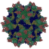
 Map data Map data | SIPV3-2E1 Complex map | |||||||||
 Sample Sample |
| |||||||||
 Keywords Keywords | Virus / Antibody / Complex | |||||||||
| Function / homology |  Function and homology information Function and homology informationcaveolin-mediated endocytosis of virus by host cell / symbiont-mediated suppression of host cytoplasmic pattern recognition receptor signaling pathway via inhibition of RIG-I activity / symbiont-mediated suppression of host cytoplasmic pattern recognition receptor signaling pathway via inhibition of MDA-5 activity / symbiont-mediated suppression of host cytoplasmic pattern recognition receptor signaling pathway via inhibition of MAVS activity / picornain 2A / symbiont-mediated suppression of host mRNA export from nucleus / symbiont genome entry into host cell via pore formation in plasma membrane / picornain 3C / T=pseudo3 icosahedral viral capsid / host cell cytoplasmic vesicle membrane ...caveolin-mediated endocytosis of virus by host cell / symbiont-mediated suppression of host cytoplasmic pattern recognition receptor signaling pathway via inhibition of RIG-I activity / symbiont-mediated suppression of host cytoplasmic pattern recognition receptor signaling pathway via inhibition of MDA-5 activity / symbiont-mediated suppression of host cytoplasmic pattern recognition receptor signaling pathway via inhibition of MAVS activity / picornain 2A / symbiont-mediated suppression of host mRNA export from nucleus / symbiont genome entry into host cell via pore formation in plasma membrane / picornain 3C / T=pseudo3 icosahedral viral capsid / host cell cytoplasmic vesicle membrane / ribonucleoside triphosphate phosphatase activity / nucleoside-triphosphate phosphatase / channel activity / monoatomic ion transmembrane transport / DNA replication / RNA helicase activity / symbiont-mediated activation of host autophagy / RNA-directed RNA polymerase / cysteine-type endopeptidase activity / viral RNA genome replication / RNA-directed RNA polymerase activity / virion attachment to host cell / host cell nucleus / DNA-templated transcription / structural molecule activity / proteolysis / RNA binding / zinc ion binding / ATP binding Similarity search - Function | |||||||||
| Biological species |  Poliovirus 3 / Poliovirus 3 /  Homo sapiens (human) Homo sapiens (human) | |||||||||
| Method | single particle reconstruction / cryo EM / Resolution: 2.95 Å | |||||||||
 Authors Authors | Waddey BT / Hafenstein SL | |||||||||
| Funding support |  United States, 1 items United States, 1 items
| |||||||||
 Citation Citation | Journal: Nat Commun / Year: 2026 Title: Neutralizing human monoclonal antibodies to poliovirus map to the receptor binding site. Authors: Benjamin T Waddey / Andrew J Charnesky / Julia E Faust / Nadia M DiNunno / Rama Devudu Puligedda / Sung Hyun Cho / Carol M Bator / Steven D Dong / Kutub Mahmood / Konstantin M Chumakov / ...Authors: Benjamin T Waddey / Andrew J Charnesky / Julia E Faust / Nadia M DiNunno / Rama Devudu Puligedda / Sung Hyun Cho / Carol M Bator / Steven D Dong / Kutub Mahmood / Konstantin M Chumakov / Scott K Dessain / Susan L Hafenstein / Abstract: Poliovirus remains a serious threat to human health. Complete eradication of wild-type poliovirus has not yet succeeded, making the development of successful antivirals critical. Microneutralization ...Poliovirus remains a serious threat to human health. Complete eradication of wild-type poliovirus has not yet succeeded, making the development of successful antivirals critical. Microneutralization assays against all three poliovirus serotypes identified a panel of human monoclonal IgGs, which are either serotype-specific or cross-neutralizing. Here, through cryoEM single particle analysis, we solved high resolution structures of four distinct poliovirus-FAb complexes. These antibodies bind to capsids at the circular depression (canyon) surrounding the icosahedral five-fold symmetry axis, which is also the binding site of the poliovirus receptor (PVR). Analysis of these structures confirms overlap of FAb contacts on the viral capsid with those of PVR. For three of the FAbs, the capsid residues are identified that dictate serotype-specific recognition. Contacts for the cross-neutralizing mAb 10D2 are located deep in the capsid canyon. These structural analyses indicate that antibody competition with the receptor likely leads to neutralization of virus particles and inhibition of poliovirus entry into host cells. Thus, the human IgGs studied here may facilitate development of therapeutics for the ongoing efforts in global eradication of poliovirus. | |||||||||
| History |
|
- Structure visualization
| Supplemental images |
|---|

- Downloads & links
-EMDB archive
| Map data | emd_70320.map.gz | 393.3 MB | EMDB map data format | |
|---|---|---|---|---|
| Header (meta data) | emd-70320-v30.xmlemd-70320.xml | 23.8 KB 23.8 KB | Display Display | EMDB header |
| FSC (resolution estimation) | emd_70320_fsc.xml | 19.8 KB | Display | FSC data file |
| Images |  emd_70320.png emd_70320.png | 61.1 KB | ||
| Filedesc metadata | emd-70320.cif.gz | 6.5 KB | ||
| Others | emd_70320_half_map_1.map.gzemd_70320_half_map_2.map.gz | 759.7 MB 759.7 MB | ||
| Archive directory |  http://ftp.pdbj.org/pub/emdb/structures/EMD-70320ftp://ftp.pdbj.org/pub/emdb/structures/EMD-70320 http://ftp.pdbj.org/pub/emdb/structures/EMD-70320ftp://ftp.pdbj.org/pub/emdb/structures/EMD-70320 | HTTPS FTP |
-Related structure data
| Related structure data |  9ocoMC  9oclC  9od3C  9oeaC M: atomic model generated by this map C: citing same article ( |
|---|---|
| Similar structure data |


-Links
| EMDB pages | EMDB (EBI/PDBe) / EMDataResource |
|---|---|
| Related items in Molecule of the Month |


-Map
| File | Download / File: emd_70320.map.gz / Format: CCP4 / Size: 824 MB / Type: IMAGE STORED AS FLOATING POINT NUMBER (4 BYTES) | ||||||||||||||||||||||||||||||||||||
|---|---|---|---|---|---|---|---|---|---|---|---|---|---|---|---|---|---|---|---|---|---|---|---|---|---|---|---|---|---|---|---|---|---|---|---|---|---|
| Annotation | SIPV3-2E1 Complex map | ||||||||||||||||||||||||||||||||||||
| Projections & slices | Image control
Images are generated by Spider. | ||||||||||||||||||||||||||||||||||||
| Voxel size | X=Y=Z: 1.08 Å | ||||||||||||||||||||||||||||||||||||
| Density |
| ||||||||||||||||||||||||||||||||||||
| Symmetry | Space group: 1 | ||||||||||||||||||||||||||||||||||||
| Details | EMDB XML:
|
 Z (Sec.)
 Y (Row.)
 X (Col.)


-Supplemental data
-Half map: Half map 1
| File | emd_70320_half_map_1.map | ||||||||||||
|---|---|---|---|---|---|---|---|---|---|---|---|---|---|
| Annotation | Half map 1 | ||||||||||||
| Projections & Slices |
| ||||||||||||
| Density Histograms |


-Half map: Half map 2
| File | emd_70320_half_map_2.map | ||||||||||||
|---|---|---|---|---|---|---|---|---|---|---|---|---|---|
| Annotation | Half map 2 | ||||||||||||
| Projections & Slices |
| ||||||||||||
| Density Histograms |


- Sample components
-Entire : SIPV3-2E1 Complex
| Entire | Name: SIPV3-2E1 Complex |
|---|---|
| Components |
|
-Supramolecule #1: SIPV3-2E1 Complex
| Supramolecule | Name: SIPV3-2E1 Complex / type: complex / ID: 1 / Parent: 0 / Macromolecule list: #1-#6 |
|---|---|
| Source (natural) | Organism: Poliovirus 3 |
| Molecular weight | Theoretical: 8 MDa |
-Macromolecule #1: VP1
| Macromolecule | Name: VP1 / type: protein_or_peptide / ID: 1 / Number of copies: 1 / Enantiomer: LEVO |
|---|---|
| Source (natural) | Organism: Poliovirus 3 |
| Molecular weight | Theoretical: 33.523672 KDa |
| Sequence | String: GIEDLISEVA QGALTLSLPK QQDSLPDTKA SGPAHSKEVP ALTAVETGAT NPLAPSDTVQ TRHVVQRRSR SESTIESFFA RGACVAIIE VDNEQPTTRA QKLFAMWRIT YKDTVQLRRK LEFFTYSRFD MEFTFVVTAN FTNANNGHAL NQVYQIMYIP P GAPTPKSW ...String: GIEDLISEVA QGALTLSLPK QQDSLPDTKA SGPAHSKEVP ALTAVETGAT NPLAPSDTVQ TRHVVQRRSR SESTIESFFA RGACVAIIE VDNEQPTTRA QKLFAMWRIT YKDTVQLRRK LEFFTYSRFD MEFTFVVTAN FTNANNGHAL NQVYQIMYIP P GAPTPKSW DDYTWQTSSN PSIFYTYGAA PARISVPYVG LANAYSHFYD GFAKVPLKTD ANDQIGDSLY SAMTVDDFGV LA VRVVNDH NPTKVTSKVR IYMKPKHVRV WCPRPPRAVP YYGPGVDYRN NLDPLSEKGL TTY UniProtKB: Genome polyprotein |
-Macromolecule #2: VP2
| Macromolecule | Name: VP2 / type: protein_or_peptide / ID: 2 / Number of copies: 1 / Enantiomer: LEVO |
|---|---|
| Source (natural) | Organism: Poliovirus 3 |
| Molecular weight | Theoretical: 30.16992 KDa |
| Sequence | String: SPNVEACGYS DRVLQLTLGN STITTQEAAN SVVAYGRWPE FIRDDEANPV DQPTEPDVAT CRFYTLDTVM WGKESKGWWW KLPDALRDM GLFGQNMYYH YLGRSGYTVH VQCNASKFHQ GALGVFAIPE YCLAGDSDKQ RYTSYANANP GERGGKFYSQ F NKDNAVTS ...String: SPNVEACGYS DRVLQLTLGN STITTQEAAN SVVAYGRWPE FIRDDEANPV DQPTEPDVAT CRFYTLDTVM WGKESKGWWW KLPDALRDM GLFGQNMYYH YLGRSGYTVH VQCNASKFHQ GALGVFAIPE YCLAGDSDKQ RYTSYANANP GERGGKFYSQ F NKDNAVTS PKREFCPVDY LLGCGVLLGN AFVYPHQIIN LRTNNSATIV LPYVNALAID SMVKHNNWGI AILPLSPLDF AQ DSSVEIP ITVTIAPMCS EFNGLRNVTA PKFQ UniProtKB: Genome polyprotein |
-Macromolecule #3: VP3
| Macromolecule | Name: VP3 / type: protein_or_peptide / ID: 3 / Number of copies: 1 / Enantiomer: LEVO |
|---|---|
| Source (natural) | Organism: Poliovirus 3 |
| Molecular weight | Theoretical: 26.289127 KDa |
| Sequence | String: GLPVLNTPGS NQYLTSDNHQ SPCAIPEFDV TPPIDIPGEV KNMMELAEID TMIPLNLEST KRNTMDMYRV TLSDSADLSQ PILCLSLSP AFDPRLSHTM LGEVLNYYTH WAGSLKFTFL FCGSMMATGK ILVAYAPPGA QPPTSRKEAM LGTHVIWDLG L QSSCTMVV ...String: GLPVLNTPGS NQYLTSDNHQ SPCAIPEFDV TPPIDIPGEV KNMMELAEID TMIPLNLEST KRNTMDMYRV TLSDSADLSQ PILCLSLSP AFDPRLSHTM LGEVLNYYTH WAGSLKFTFL FCGSMMATGK ILVAYAPPGA QPPTSRKEAM LGTHVIWDLG L QSSCTMVV PWISNVTYRQ TTQDSFTEGG YISMFYQTRI VVPLSTPKSM SMLGFVSACN DFSVRLLRDT THISQSALPQ UniProtKB: Genome polyprotein |
-Macromolecule #4: VP4
| Macromolecule | Name: VP4 / type: protein_or_peptide / ID: 4 / Number of copies: 1 / Enantiomer: LEVO |
|---|---|
| Source (natural) | Organism: Poliovirus 3 |
| Molecular weight | Theoretical: 7.320917 KDa |
| Sequence | String: GAQVSSQKVG AHENSNRAYG GSTINYTTIN YYKDSASNAA SKQDYSQDPS KFTEPLKDVL IKTAPALN UniProtKB: Genome polyprotein |
-Macromolecule #5: 2E1 Heavy Chain
| Macromolecule | Name: 2E1 Heavy Chain / type: protein_or_peptide / ID: 5 / Number of copies: 1 / Enantiomer: LEVO |
|---|---|
| Source (natural) | Organism: Homo sapiens (human) |
| Molecular weight | Theoretical: 14.006592 KDa |
| Sequence | String: EVQLVQSGAE VKKSGESLKI SCKASGYHFT SYWIGWVRQM PGKGLEWMGF IYPGDSDTRY GPSFQGQVSI SADKSSSTAY LQWSRLKAS DTAMYFCARH GGFASSLYSS SFRPPDYWGQ GTLVTVSS |
-Macromolecule #6: 2E1 Light Chain
| Macromolecule | Name: 2E1 Light Chain / type: protein_or_peptide / ID: 6 / Number of copies: 1 / Enantiomer: LEVO |
|---|---|
| Source (natural) | Organism: Homo sapiens (human) |
| Molecular weight | Theoretical: 11.837788 KDa |
| Sequence | String: NFMLTQPHSV SESPGKTVTI SCTRSSGSIA SSYVQWYQQR PGSSPTTVIY EDNQRPSGVP DRFSGSIDSS SNSASLIISG LKTEDEADY YCQSYDSSNQ VFGGGTKLTV L |
-Macromolecule #7: PALMITIC ACID
| Macromolecule | Name: PALMITIC ACID / type: ligand / ID: 7 / Number of copies: 1 / Formula: PLM |
|---|---|
| Molecular weight | Theoretical: 256.424 Da |
| Chemical component information |  ChemComp-PLM: |
-Experimental details
-Structure determination
| Method | cryo EM |
|---|---|
 Processing Processing | single particle reconstruction |
| Aggregation state | particle |
-Sample preparation
| Buffer | pH: 8 |
|---|---|
| Vitrification | Cryogen name: ETHANE |
- Electron microscopy
| Microscope | TFS KRIOS |
|---|---|
| Image recording | Film or detector model: GATAN K3 (6k x 4k) / Average electron dose: 56.0 e/Å2 |
| Electron beam | Acceleration voltage: 300 kV / Electron source:  FIELD EMISSION GUN FIELD EMISSION GUN |
| Electron optics | Illumination mode: FLOOD BEAM / Imaging mode: BRIGHT FIELD / Nominal defocus max: 2.5 µm / Nominal defocus min: 0.5 µm |
| Experimental equipment |  Model: Titan Krios / Image courtesy: FEI Company |