 Movie
Movie Controller

[English] 日本語
 Yorodumi
- EMDB-70051: Structure of WT E.coli ribosome 70S subunit with complexed with m... -
+ Open data

- Basic information
| Entry |  | |||||||||
|---|---|---|---|---|---|---|---|---|---|---|
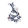
| Title | Structure of WT E.coli ribosome 70S subunit with complexed with mRNA, P-site fMet-NH-tRNAfMet and A-site (R) beta-2-hydroxy-BocLysine acid charged NH-tRNAPyl | |||||||||
 Map data Map data | composite 70S map | |||||||||
 Sample Sample |
| |||||||||
 Keywords Keywords | ribosome / non-natural monomers / beta-2-hydroxy acids / unnatural monomers | |||||||||
| Function / homology |  Function and homology information Function and homology informationnegative regulation of cytoplasmic translational initiation / transcriptional attenuation / endoribonuclease inhibitor activity / RNA-binding transcription regulator activity / negative regulation of cytoplasmic translation / DnaA-L2 complex / translation repressor activity / negative regulation of translational initiation / negative regulation of DNA-templated DNA replication initiation / mRNA regulatory element binding translation repressor activity ...negative regulation of cytoplasmic translational initiation / transcriptional attenuation / endoribonuclease inhibitor activity / RNA-binding transcription regulator activity / negative regulation of cytoplasmic translation / DnaA-L2 complex / translation repressor activity / negative regulation of translational initiation / negative regulation of DNA-templated DNA replication initiation / mRNA regulatory element binding translation repressor activity / ribosome assembly / assembly of large subunit precursor of preribosome / transcription antitermination / translational initiation / DNA-templated transcription termination / response to radiation / maintenance of translational fidelity / mRNA 5'-UTR binding / large ribosomal subunit / transferase activity / ribosomal small subunit assembly / ribosome binding / ribosomal small subunit biogenesis / 5S rRNA binding / small ribosomal subunit / ribosomal large subunit assembly / small ribosomal subunit rRNA binding / cytosolic small ribosomal subunit / large ribosomal subunit rRNA binding / cytosolic large ribosomal subunit / cytoplasmic translation / tRNA binding / negative regulation of translation / rRNA binding / structural constituent of ribosome / ribosome / translation / ribonucleoprotein complex / response to antibiotic / negative regulation of DNA-templated transcription / mRNA binding / DNA binding / RNA binding / zinc ion binding / membrane / cytoplasm / cytosol Similarity search - Function | |||||||||
| Biological species |   Methanomethylophilus alvi (archaea) Methanomethylophilus alvi (archaea) | |||||||||
| Method | single particle reconstruction / cryo EM / Resolution: 2.14 Å | |||||||||
 Authors Authors | Majumdar C / Cate JHD | |||||||||
| Funding support |  United States, 1 items United States, 1 items
| |||||||||
 Citation Citation | Journal: J Am Chem Soc / Year: 2026 Title: Co-Translational Incorporation of - and -β-Hydroxy Acids : A Structural and Biochemical Study on the Ribosome. Authors: Chandrima Majumdar / Alexandra D Kent / Noah X Hamlish / Cathy Zhu / Katelyn A Fitzgerald / Jamie H D Cate / Alanna Schepartz / Abstract: Engineering the translation apparatus to accept backbone-modified amino acid analogues would enable the programmed synthesis of sequence-defined biopolymers with tunable properties. β-Hydroxy acids ...Engineering the translation apparatus to accept backbone-modified amino acid analogues would enable the programmed synthesis of sequence-defined biopolymers with tunable properties. β-Hydroxy acids are of particular interest because they could support the programmed biosynthesis of both biocompatible polyester materials as well as natural product-like depsipeptides. Previous work has reported that both enantiomers of β-hydroxy-N-Boc-lysine (β-OH-BocK) are substrates for the orthogonal pyrrolysyl-tRNA synthetase (PylRS)/tRNA pair, but only one enantiomer is introduced into protein . Here we make use of high-resolution cryogenic electron microscopy (cryo-EM) to determine the structural basis for this observation. These structures reveal both β-OH-BocK isomers equally well-positioned within the ribosomal A site regardless of stereochemistry. Consistent with this observation, translation reactions charged with tRNAs acylated with - or -β-OH-BocK produced roughly equal amounts of translated product when quantified on the basis of either mass spectrometry or luminescence. Together, these experiments imply that the substantial preferential incorporation of one enantiomer over the other observed previously results primarily from deficiencies in the steps that precede bond formation by the ribosome. Indeed, as predicted by this work and demonstrated in an accompanying paper (Soni, C. Co-Translational Incorporation of ()- and ()-β-Hydroxyacids : Directed Evolution of Efficient Aminoacyl-tRNA Synthetases. 2026, 148, 10.1021/jacs.5c18595), when cells are provided with an active and orthogonal aminoacyl-tRNA synthetase/tRNA pair that accepts both - and -β-OH-BocK as substrates, both monomers are introduced into protein in good yield and with high fidelity. | |||||||||
| History |
|
- Structure visualization
| Supplemental images |
|---|

- Downloads & links
-EMDB archive
| Map data | emd_70051.map.gz | 63.6 MB | EMDB map data format | |
|---|---|---|---|---|
| Header (meta data) | emd-70051-v30.xmlemd-70051.xml | 73.7 KB 73.7 KB | Display Display | EMDB header |
| FSC (resolution estimation) | emd_70051_fsc.xml | 18 KB | Display | FSC data file |
| Images |  emd_70051.png emd_70051.png | 85.2 KB | ||
| Filedesc metadata | emd-70051.cif.gz | 15.6 KB | ||
| Archive directory |  http://ftp.pdbj.org/pub/emdb/structures/EMD-70051ftp://ftp.pdbj.org/pub/emdb/structures/EMD-70051 http://ftp.pdbj.org/pub/emdb/structures/EMD-70051ftp://ftp.pdbj.org/pub/emdb/structures/EMD-70051 | HTTPS FTP |
-Related structure data
| Related structure data |  9o2yMC  9o2xC C: citing same article ( M: atomic model generated by this map |
|---|---|
| Similar structure data |


-Links
| EMDB pages | EMDB (EBI/PDBe) / EMDataResource |
|---|---|
| Related items in Molecule of the Month |


-Map
| File | Download / File: emd_70051.map.gz / Format: CCP4 / Size: 512 MB / Type: IMAGE STORED AS FLOATING POINT NUMBER (4 BYTES) | ||||||||||||||||||||||||||||||||||||
|---|---|---|---|---|---|---|---|---|---|---|---|---|---|---|---|---|---|---|---|---|---|---|---|---|---|---|---|---|---|---|---|---|---|---|---|---|---|
| Annotation | composite 70S map | ||||||||||||||||||||||||||||||||||||
| Projections & slices | Image control
Images are generated by Spider. | ||||||||||||||||||||||||||||||||||||
| Voxel size | X=Y=Z: 0.8235 Å | ||||||||||||||||||||||||||||||||||||
| Density |
| ||||||||||||||||||||||||||||||||||||
| Symmetry | Space group: 1 | ||||||||||||||||||||||||||||||||||||
| Details | EMDB XML:
|
 Z (Sec.)
 Y (Row.)
 X (Col.)


-Supplemental data
- Sample components
+Entire : 70S ribosome particle
+Supramolecule #1: 70S ribosome particle
+Macromolecule #1: 16S rRNA
+Macromolecule #22: mRNA
+Macromolecule #23: P-site tRNA fMet
+Macromolecule #24: 23S rRNA
+Macromolecule #25: 5S rRNA
+Macromolecule #55: A-site tRNAPyl
+Macromolecule #2: 30S ribosomal protein S2
+Macromolecule #3: Small ribosomal subunit protein uS3
+Macromolecule #4: Small ribosomal subunit protein uS4
+Macromolecule #5: Small ribosomal subunit protein uS5
+Macromolecule #6: Small ribosomal subunit protein bS6
+Macromolecule #7: Small ribosomal subunit protein uS7
+Macromolecule #8: Small ribosomal subunit protein uS8
+Macromolecule #9: Small ribosomal subunit protein uS9
+Macromolecule #10: Small ribosomal subunit protein uS10
+Macromolecule #11: Small ribosomal subunit protein uS11
+Macromolecule #12: Small ribosomal subunit protein uS12
+Macromolecule #13: Small ribosomal subunit protein uS13
+Macromolecule #14: Small ribosomal subunit protein uS14
+Macromolecule #15: Small ribosomal subunit protein uS15
+Macromolecule #16: Small ribosomal subunit protein bS16
+Macromolecule #17: Small ribosomal subunit protein uS17
+Macromolecule #18: Small ribosomal subunit protein bS18
+Macromolecule #19: Small ribosomal subunit protein uS19
+Macromolecule #20: Small ribosomal subunit protein bS20
+Macromolecule #21: Small ribosomal subunit protein bS21
+Macromolecule #26: 50S ribosomal protein L2
+Macromolecule #27: 50S ribosomal protein L3
+Macromolecule #28: Large ribosomal subunit protein uL4
+Macromolecule #29: Large ribosomal subunit protein uL5
+Macromolecule #30: Large ribosomal subunit protein uL6
+Macromolecule #31: Large ribosomal subunit protein bL9
+Macromolecule #32: Large ribosomal subunit protein uL13
+Macromolecule #33: Large ribosomal subunit protein uL14
+Macromolecule #34: Large ribosomal subunit protein uL15
+Macromolecule #35: 50S ribosomal protein L16
+Macromolecule #36: Large ribosomal subunit protein bL17
+Macromolecule #37: Large ribosomal subunit protein uL18
+Macromolecule #38: Large ribosomal subunit protein bL19
+Macromolecule #39: 50S ribosomal protein L20
+Macromolecule #40: Large ribosomal subunit protein bL21
+Macromolecule #41: Large ribosomal subunit protein uL22
+Macromolecule #42: 50S ribosomal protein L23
+Macromolecule #43: 50S ribosomal protein L24
+Macromolecule #44: Large ribosomal subunit protein bL25
+Macromolecule #45: Large ribosomal subunit protein bL27
+Macromolecule #46: 50S ribosomal protein L28
+Macromolecule #47: Large ribosomal subunit protein uL29
+Macromolecule #48: 50S ribosomal protein L30
+Macromolecule #49: 50S ribosomal protein L32
+Macromolecule #50: 50S ribosomal protein L33
+Macromolecule #51: 50S ribosomal protein L34
+Macromolecule #52: 50S ribosomal protein L35
+Macromolecule #53: 50S ribosomal protein L36
+Macromolecule #54: 50S ribosomal protein L31
+Macromolecule #56: PAROMOMYCIN
+Macromolecule #57: MAGNESIUM ION
+Macromolecule #58: SPERMIDINE

+Macromolecule #59: N-FORMYLMETHIONINE
+Macromolecule #60: SPERMINE

+Macromolecule #61: POTASSIUM ION
+Macromolecule #62: ZINC ION
+Macromolecule #63: (2R)-6-[(tert-butoxycarbonyl)amino]-2-(hydroxymethyl)hexanoic acid
+Macromolecule #64: water

-Experimental details
-Structure determination
| Method | cryo EM |
|---|---|
 Processing Processing | single particle reconstruction |
| Aggregation state | particle |
-Sample preparation
| Buffer | pH: 7.5 |
|---|---|
| Grid | Model: Quantifoil R1.2/1.3 / Material: GOLD / Mesh: 300 / Support film - Material: CARBON / Support film - topology: CONTINUOUS / Support film - Film thickness: 2 / Pretreatment - Type: GLOW DISCHARGE / Pretreatment - Time: 12 sec. |
| Vitrification | Cryogen name: ETHANE / Chamber humidity: 100 % / Chamber temperature: 277 K / Instrument: FEI VITROBOT MARK IV |
- Electron microscopy
| Microscope | TFS KRIOS |
|---|---|
| Image recording | Film or detector model: GATAN K3 BIOQUANTUM (6k x 4k) / Average electron dose: 40.0 e/Å2 |
| Electron beam | Acceleration voltage: 300 kV / Electron source:  FIELD EMISSION GUN FIELD EMISSION GUN |
| Electron optics | Illumination mode: OTHER / Imaging mode: BRIGHT FIELD / Nominal defocus max: 1.5 µm / Nominal defocus min: 0.5 µm |
| Experimental equipment |  Model: Titan Krios / Image courtesy: FEI Company |
+Image processing

-Atomic model buiding 1
| Initial model | PDB ID: Chain - Source name: PDB / Chain - Initial model type: experimental model |
|---|---|
| Refinement | Protocol: RIGID BODY FIT |
| Output model | PDB-9o2y: |
