Movie
Movie Controller

[English] 日本語
 Yorodumi
- EMDB-65579: Cryo-EM structure of Fo domain of FoF1-ATPase monomer state on th... -
+ Open data

- Basic information
| Entry |  | ||||||||||||
|---|---|---|---|---|---|---|---|---|---|---|---|---|---|
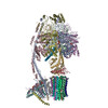
| Title | Cryo-EM structure of Fo domain of FoF1-ATPase monomer state on the bovine heart submitochondrial particles | ||||||||||||
 Map data Map data | |||||||||||||
 Sample Sample |
| ||||||||||||
 Keywords Keywords | ATP synthase / FoF1-ATPase / submitochondrial particles / MOTOR PROTEIN | ||||||||||||
| Function / homology |  Function and homology information Function and homology informationMitochondrial protein import / Formation of ATP by chemiosmotic coupling / Cristae formation / Mitochondrial translation termination / proton channel activity / Mitochondrial protein degradation / proton transmembrane transporter activity / proton motive force-driven ATP synthesis / proton-transporting two-sector ATPase complex, proton-transporting domain / proton motive force-driven mitochondrial ATP synthesis ...Mitochondrial protein import / Formation of ATP by chemiosmotic coupling / Cristae formation / Mitochondrial translation termination / proton channel activity / Mitochondrial protein degradation / proton transmembrane transporter activity / proton motive force-driven ATP synthesis / proton-transporting two-sector ATPase complex, proton-transporting domain / proton motive force-driven mitochondrial ATP synthesis / proton-transporting ATP synthase complex / proton-transporting ATP synthase activity, rotational mechanism / proton transmembrane transport / mitochondrial membrane / mitochondrial inner membrane / lipid binding / mitochondrion Similarity search - Function | ||||||||||||
| Biological species |  | ||||||||||||
| Method | single particle reconstruction / cryo EM / Resolution: 4.1 Å | ||||||||||||
 Authors Authors | Nakano A / Masuya T / Akisada S / Ishikawa-Fukuda M / Mitsuoka K / Miyoshi H / Murai M / Yokoyama K | ||||||||||||
| Funding support |  Japan, 3 items Japan, 3 items
| ||||||||||||
 Citation Citation | Journal: Nat Commun / Year: 2026 Title: Structures of respiratory supercomplexes and ATP synthase oligomers in mammalian mitochondrial inner membrane. Authors: Atsuki Nakano / Takahiro Masuya / Shinsuke Akisada / Moe Ishikawa-Fukuda / Kaoru Mitsuoka / Hideto Miyoshi / Masatoshi Murai / Ken Yokoyama / Abstract: Understanding the functional mechanisms of membrane protein complexes requires structural analysis within their native membrane environment. Here, we applied cryo-electron microscopy to determine the ...Understanding the functional mechanisms of membrane protein complexes requires structural analysis within their native membrane environment. Here, we applied cryo-electron microscopy to determine the structures of FF ATP synthase and respiratory supercomplexes (SCs) on sub-mitochondrial particles (SMPs) isolated from bovine heart mitochondria. Most FF complexes were observed as dimers stabilized by the regulatory factor IF₁, and a tetrameric assembly comprising two FF-IF₁ dimers arranged linearly was also identified. This finding indicates that the tetrameric units of FF are present in the mitochondrial inner membrane and contribute to shaping cristae tips in mammalian mitochondria. F domain maps resolve the e-subunit- c₈-ring interface and show no discrete density for a tightly bound lipid within the c₈-ring. In addition to the previously reported SCs compositions CI₁CIII₂CIV₁ and CI₁CIII₂CIV₂, our analysis identified an additional assembly with the composition CI₁CIII₂CIV₃, as well as a CI₂CIII₂CIV₆ mega-complex. This approach enables rapid structural determination of FF ATP synthase and SCs from minimal membrane fractions, providing a foundation for elucidating the molecular basis of metabolic disorders and mitochondrial diseases at the level of higher-order architecture. | ||||||||||||
| History |
|
- Structure visualization
| Supplemental images |
|---|

- Downloads & links
-EMDB archive
| Map data | emd_65579.map.gz | 328.1 MB | EMDB map data format | |
|---|---|---|---|---|
| Header (meta data) | emd-65579-v30.xmlemd-65579.xml | 26.9 KB 26.9 KB | Display Display | EMDB header |
| FSC (resolution estimation) | emd_65579_fsc.xml | 14.9 KB | Display | FSC data file |
| Images |  emd_65579.png emd_65579.png | 135 KB | ||
| Masks | emd_65579_msk_1.map | 347.6 MB | Mask map | |
| Filedesc metadata | emd-65579.cif.gz | 6.8 KB | ||
| Others | emd_65579_half_map_1.map.gzemd_65579_half_map_2.map.gz | 322.3 MB 322.3 MB | ||
| Archive directory |  http://ftp.pdbj.org/pub/emdb/structures/EMD-65579ftp://ftp.pdbj.org/pub/emdb/structures/EMD-65579 http://ftp.pdbj.org/pub/emdb/structures/EMD-65579ftp://ftp.pdbj.org/pub/emdb/structures/EMD-65579 | HTTPS FTP |
-Related structure data
| Related structure data |  9w2tMC  9w2rC  9w2sC  9w2uC  9w2vC  9w2xC  9w2yC  9w2zC M: atomic model generated by this map C: citing same article ( |
|---|---|
| Similar structure data |


-Links
| EMDB pages | EMDB (EBI/PDBe) / EMDataResource |
|---|---|
| Related items in Molecule of the Month |


-Map
| File | Download / File: emd_65579.map.gz / Format: CCP4 / Size: 347.6 MB / Type: IMAGE STORED AS FLOATING POINT NUMBER (4 BYTES) | ||||||||||||||||||||||||||||||||||||
|---|---|---|---|---|---|---|---|---|---|---|---|---|---|---|---|---|---|---|---|---|---|---|---|---|---|---|---|---|---|---|---|---|---|---|---|---|---|
| Projections & slices | Image control
Images are generated by Spider. | ||||||||||||||||||||||||||||||||||||
| Voxel size | X=Y=Z: 1.45333 Å | ||||||||||||||||||||||||||||||||||||
| Density |
| ||||||||||||||||||||||||||||||||||||
| Symmetry | Space group: 1 | ||||||||||||||||||||||||||||||||||||
| Details | EMDB XML:
|
 Z (Sec.)
 Y (Row.)
 X (Col.)


-Supplemental data
-Mask #1
| File | emd_65579_msk_1.map | ||||||||||||
|---|---|---|---|---|---|---|---|---|---|---|---|---|---|
| Projections & Slices |
| ||||||||||||
| Density Histograms |


-Half map: #2
| File | emd_65579_half_map_1.map | ||||||||||||
|---|---|---|---|---|---|---|---|---|---|---|---|---|---|
| Projections & Slices |
| ||||||||||||
| Density Histograms |


-Half map: #1
| File | emd_65579_half_map_2.map | ||||||||||||
|---|---|---|---|---|---|---|---|---|---|---|---|---|---|
| Projections & Slices |
| ||||||||||||
| Density Histograms |


- Sample components
+Entire : FoF1-ATPase on the bovine heart submitochondrial particles
+Supramolecule #1: FoF1-ATPase on the bovine heart submitochondrial particles
+Macromolecule #1: ATP synthase F(0) complex subunit 8
+Macromolecule #2: ATP synthase F(0) complex subunit C1, mitochondrial
+Macromolecule #3: ATP synthase F(0) complex subunit a
+Macromolecule #4: ATP synthase peripheral stalk subunit b, mitochondrial
+Macromolecule #5: ATP synthase peripheral stalk subunit d, mitochondrial
+Macromolecule #6: ATP synthase F(0) complex subunit e, mitochondrial
+Macromolecule #7: ATP synthase F(0) complex subunit f, mitochondrial
+Macromolecule #8: ATP synthase F(0) complex subunit g, mitochondrial
+Macromolecule #9: ATP synthase F(0) complex subunit j, mitochondrial
+Macromolecule #10: ATP synthase F(0) complex subunit k, mitochondrial
-Experimental details
-Structure determination
| Method | cryo EM |
|---|---|
 Processing Processing | single particle reconstruction |
| Aggregation state | particle |
-Sample preparation
| Buffer | pH: 7.5 |
|---|---|
| Vitrification | Cryogen name: ETHANE / Chamber humidity: 100 % / Chamber temperature: 298 K |
- Electron microscopy
| Microscope | TFS KRIOS |
|---|---|
| Image recording | Film or detector model: GATAN K3 (6k x 4k) / Average electron dose: 50.0 e/Å2 |
| Electron beam | Acceleration voltage: 300 kV / Electron source:  FIELD EMISSION GUN FIELD EMISSION GUN |
| Electron optics | Illumination mode: FLOOD BEAM / Imaging mode: BRIGHT FIELD / Cs: 2.7 mm / Nominal defocus max: 1.8 µm / Nominal defocus min: 0.8 µm |
| Experimental equipment |  Model: Titan Krios / Image courtesy: FEI Company |